Ranica Arrowsmith, Associate Editor05.06.15
Ocular technology doesn’t usually get a lot of attention, as visual impairments aren’t as sexy as, say. cardio or neurotech, which often address life-threatening conditions. However, as the global medical market faces the problem of a growing aging population, visual ailments such as cataracts warrant further attention. Other ocular diseases, such as Leber’s hereditary optic neuropathy or anophthalmia, are classified as rare, and as such are not as attractive for research and development funding as many of the more common diseases.
According to the most recent data from Prevent Blindness America, a volunteer eye health and safety organization, there has been an alarming rise in ocular disease. A rise in diabetes has led to an 89 percent rise in diabetic retinopathy over the 12 years prior to the report. Macular degeneration was up 25 percent, due to a larger elderly population.
In the United States, according to the Centers for Disease Control and Prevention, 2 million Americans older than the age of 50 have age-related macular degeneration; 22 million Americans over the age of 40 have cataracts; 4.5 million Americans older than 40 have diabetic retinopathy; and 2.3 million Americans older than 40 have glaucoma.
The biological and chemical structure of the eye makes it especially impervious to drug intervention, which is why the medical device industry is so important to addressing the varied needs of the ocular disease space.1
Highlighted below are two new clearances from the U.S. Food and Drug Administration (FDA) that address the ocular surgical space, both of which address a wide range of ophthalmological needs as opposed to a narrow, specialized target.
The first approval is a surgical microscope from Carl Zeiss Meditec AG, which can be used for a number of surgical procedures concerning the eye, including complex detachments necessitated by proliferative diabetic retinopathy and proliferative vitreoretinopathy.
The second is a clearance for five application upgrades to Lensar’s laser system, which targets cataracts.
FDA Clears New Surgical Microscope for Eye Surgery
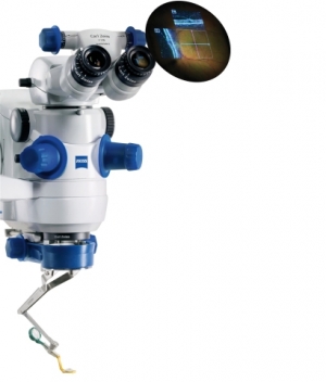
The FDA has cleared for sale a new surgical microscope from Carl Zeiss Meditec that helps improve the imaging of certain anatomical details during eye surgery.
High-definition optical coherence tomography (OCT) images appear directly in the Lumera 700 microscope eyepiece, adding a real-time third dimension to visualization capabilities. Surgeons are provided improved views below the surface of the surgical field—enabling them to see more, even transparent structures in the anterior and posterior segment of the eye, the company said.
“Intraoperative OCT allows me to see things I could not see otherwise,” said Justis P. Ehlers, M.D., of Cleveland, Ohio, who has been using Rescan 700 on an investigational basis for nine months. “I use intraoperative OCT in most of my surgeries. We have found that real-time OCT feedback during surgery can improve our understanding of anatomy and impact surgical decision-making, particularly in membrane peeling cases and complex detachments—for example, proliferative diabetic retinopathy and proliferative vitreoretinopathy.”
The Rescan 700 helps surgeons concentrate more on the surgical procedure, as the necessary structural information known from pre-operative OCTs constantly is available intraoperatively. Additionally, the continuous OCT scanning supports achieving better patient outcomes because the surgeon can monitor progress and verify results during the procedure, Zeiss bigwigs noted in a news release.
“One of the great features of the Rescan 700 is the foot pedal control of the OCT beam—I can rapidly place it right where I need it during the surgery,” said Ehlers, the lead investigator for a prospective study with more than 200 cases examining microscope-integrated OCT and ophthalmic surgery. “And the Z-tracking feature enables improved stability of the OCT image during surgery-induced motion.”
OCT scans also can be stored and recalled for review. That way the new device enables better decision-making during surgery. “As a surgeon, I appreciate the confirmation of my surgical objectives that intraoperative OCT provides,” Ehlers said. “I receive guidance for where it’s optimal to start surgical maneuvers, and then subsequent feedback on whether I have accomplished the surgical objectives.”
Rescan 700 is useful for various surgeries in the anterior and posterior segment of the eye with a broad range of potential applications, the company noted.
“We are happy that now U.S. surgeons can also benefit from this new visualization technology and its potential to help them improve patient outcomes,” said Ludwin Monz, Ph.D., president and CEO of Carl Zeiss Meditec. “The merging of two of our gold standards into one system creates vast possibilities for changing ophthalmic surgery and expanding the capabilities of surgeons, especially for retina and cornea procedures.”
Carl Zeiss Meditec AG, listed on TecDAX of the German stock exchange, develops eye disease diagnosis and treatment technologies, including implants and consumable materials. Based in Jena, Germany, the company operates subsidiaries in Germany and at least a half-dozen foreign countries (China, France, India, Japan, Spain and the United States). Roughly 65 percent of Carl Zeiss Meditec shares are held by Carl Zeiss AG, a wholly owned subsidiary of the Carl Zeiss Foundation.
New Ensuite Technologies for Lensar Laser System
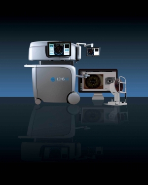
Orlando, Fla.-based Lensar Inc., which makes femtosecond laser technology for refractive cataract surgery, has received 510(k) clearance from the FDA for a suite of five new application technologies integrated into the company’s flagship product, the Lensar laser system. The Lensar laser system with Streamline is a femtosecond laser cataract platform to enable automation of several surgical procedure planning and execution elements with the introduction of five new application upgrades that include: wireless integration with the Cassini Corneal Shape Analyzer; iris registration; cataract density imaging; customized fragmentation patterns; and arcuate incision planning.
“The addition of the Streamline advanced technology suite solidifies our Lensar laser system as the femtosecond platform of choice for today’s refractive cataract surgeon,” said CEO Nicholas T. Curtis. “The five distinct upgrades to the platform include innovative integration of functions unique to the Lensar Laser System that, for the first time, allow surgeons to fully automate and customize critical planning and execution steps of refractive cataract surgery.”
According to the company, its Lensar laser system is the first femtosecond laser cataract platform to establish a wireless integration protocol with preoperative diagnostic devices. Lensar is initiating this capability with the Cassini Corneal Shape Analyzer, enabling the wireless transfer of data from preoperative corneal measurements to the Lensar laser system. This new integration is hoped to eliminate potential errors that can occur from manual entry of data from the device used in the preoperative and surgical planning process. The integration with Cassini enables the capture of preoperative iris registration data, and maps it to the image of the eye obtained under the laser at procedure time, eliminating the need to manually ink mark the eye to identify and adjust for the cyclorotation that may occur when a patient is reclined during surgery.
“Creating an integrated technology approach to the femtosecond cataract procedure is something we refractive cataract surgeons have been waiting for, and it doesn’t surprise me at all that Lensar was the company to get it done,” said Jonathan Solomon, M.D., surgical/refractive director at Solomon Eye Associates in Maryland. “I anticipate Lensar’s Streamline will improve not only O.R. (operating room) efficiency and patient experience during the procedure, but may also improve outcomes by minimizing the human factor inherent in manual data input or physically marking the eye pre-operatively.”
Additionally, surgeons have access to an arcuate (curved) incision planning table on the laser that includes parameters to define the location, depth and extent of the surgeon’s intended arcuate incisions based on individual patient biometric measurements and other factors defined by the surgeon. The arcuate incision planning capability allows surgeons to retain their plan preferences for later use, increasing surgical operating room efficiency.
The Streamline upgrades also introduce integrated cataract density imaging, which automatically categorizes the cataract to a pre-programmed, surgeon-customized fragmentation pattern depending upon the density of the cataract and allows the surgeon to automatically isolate fragmentation to the nucleus. This first-in-market innovation may have a positive impact on efficiency and procedure time, the company claims.
“Lensar has given the reigns to the surgeon to make the laser-assisted refractive cataract procedure customized to both patient and surgeon needs,” said Robert Weinstock, M.D., director of cataract and refractive services at the Eye Institute of West Florida and The Weinstock Laser Eye Center. “The ability to preprogram the laser just one time with a surgeon’s specific preferences for fragmentation patterns based upon an automated cataract density analysis and customized arcuate incision plans may increase the efficiency and accuracy for every procedure thereafter, enhancing patient comfort and, ultimately, visual outcomes.”
Reference
According to the most recent data from Prevent Blindness America, a volunteer eye health and safety organization, there has been an alarming rise in ocular disease. A rise in diabetes has led to an 89 percent rise in diabetic retinopathy over the 12 years prior to the report. Macular degeneration was up 25 percent, due to a larger elderly population.
In the United States, according to the Centers for Disease Control and Prevention, 2 million Americans older than the age of 50 have age-related macular degeneration; 22 million Americans over the age of 40 have cataracts; 4.5 million Americans older than 40 have diabetic retinopathy; and 2.3 million Americans older than 40 have glaucoma.
The biological and chemical structure of the eye makes it especially impervious to drug intervention, which is why the medical device industry is so important to addressing the varied needs of the ocular disease space.1
Highlighted below are two new clearances from the U.S. Food and Drug Administration (FDA) that address the ocular surgical space, both of which address a wide range of ophthalmological needs as opposed to a narrow, specialized target.
The first approval is a surgical microscope from Carl Zeiss Meditec AG, which can be used for a number of surgical procedures concerning the eye, including complex detachments necessitated by proliferative diabetic retinopathy and proliferative vitreoretinopathy.
The second is a clearance for five application upgrades to Lensar’s laser system, which targets cataracts.
FDA Clears New Surgical Microscope for Eye Surgery
The FDA has cleared for sale a new surgical microscope from Carl Zeiss Meditec that helps improve the imaging of certain anatomical details during eye surgery.
High-definition optical coherence tomography (OCT) images appear directly in the Lumera 700 microscope eyepiece, adding a real-time third dimension to visualization capabilities. Surgeons are provided improved views below the surface of the surgical field—enabling them to see more, even transparent structures in the anterior and posterior segment of the eye, the company said.
“Intraoperative OCT allows me to see things I could not see otherwise,” said Justis P. Ehlers, M.D., of Cleveland, Ohio, who has been using Rescan 700 on an investigational basis for nine months. “I use intraoperative OCT in most of my surgeries. We have found that real-time OCT feedback during surgery can improve our understanding of anatomy and impact surgical decision-making, particularly in membrane peeling cases and complex detachments—for example, proliferative diabetic retinopathy and proliferative vitreoretinopathy.”
The Rescan 700 helps surgeons concentrate more on the surgical procedure, as the necessary structural information known from pre-operative OCTs constantly is available intraoperatively. Additionally, the continuous OCT scanning supports achieving better patient outcomes because the surgeon can monitor progress and verify results during the procedure, Zeiss bigwigs noted in a news release.
“One of the great features of the Rescan 700 is the foot pedal control of the OCT beam—I can rapidly place it right where I need it during the surgery,” said Ehlers, the lead investigator for a prospective study with more than 200 cases examining microscope-integrated OCT and ophthalmic surgery. “And the Z-tracking feature enables improved stability of the OCT image during surgery-induced motion.”
OCT scans also can be stored and recalled for review. That way the new device enables better decision-making during surgery. “As a surgeon, I appreciate the confirmation of my surgical objectives that intraoperative OCT provides,” Ehlers said. “I receive guidance for where it’s optimal to start surgical maneuvers, and then subsequent feedback on whether I have accomplished the surgical objectives.”
Rescan 700 is useful for various surgeries in the anterior and posterior segment of the eye with a broad range of potential applications, the company noted.
“We are happy that now U.S. surgeons can also benefit from this new visualization technology and its potential to help them improve patient outcomes,” said Ludwin Monz, Ph.D., president and CEO of Carl Zeiss Meditec. “The merging of two of our gold standards into one system creates vast possibilities for changing ophthalmic surgery and expanding the capabilities of surgeons, especially for retina and cornea procedures.”
Carl Zeiss Meditec AG, listed on TecDAX of the German stock exchange, develops eye disease diagnosis and treatment technologies, including implants and consumable materials. Based in Jena, Germany, the company operates subsidiaries in Germany and at least a half-dozen foreign countries (China, France, India, Japan, Spain and the United States). Roughly 65 percent of Carl Zeiss Meditec shares are held by Carl Zeiss AG, a wholly owned subsidiary of the Carl Zeiss Foundation.
New Ensuite Technologies for Lensar Laser System
Orlando, Fla.-based Lensar Inc., which makes femtosecond laser technology for refractive cataract surgery, has received 510(k) clearance from the FDA for a suite of five new application technologies integrated into the company’s flagship product, the Lensar laser system. The Lensar laser system with Streamline is a femtosecond laser cataract platform to enable automation of several surgical procedure planning and execution elements with the introduction of five new application upgrades that include: wireless integration with the Cassini Corneal Shape Analyzer; iris registration; cataract density imaging; customized fragmentation patterns; and arcuate incision planning.
“The addition of the Streamline advanced technology suite solidifies our Lensar laser system as the femtosecond platform of choice for today’s refractive cataract surgeon,” said CEO Nicholas T. Curtis. “The five distinct upgrades to the platform include innovative integration of functions unique to the Lensar Laser System that, for the first time, allow surgeons to fully automate and customize critical planning and execution steps of refractive cataract surgery.”
According to the company, its Lensar laser system is the first femtosecond laser cataract platform to establish a wireless integration protocol with preoperative diagnostic devices. Lensar is initiating this capability with the Cassini Corneal Shape Analyzer, enabling the wireless transfer of data from preoperative corneal measurements to the Lensar laser system. This new integration is hoped to eliminate potential errors that can occur from manual entry of data from the device used in the preoperative and surgical planning process. The integration with Cassini enables the capture of preoperative iris registration data, and maps it to the image of the eye obtained under the laser at procedure time, eliminating the need to manually ink mark the eye to identify and adjust for the cyclorotation that may occur when a patient is reclined during surgery.
“Creating an integrated technology approach to the femtosecond cataract procedure is something we refractive cataract surgeons have been waiting for, and it doesn’t surprise me at all that Lensar was the company to get it done,” said Jonathan Solomon, M.D., surgical/refractive director at Solomon Eye Associates in Maryland. “I anticipate Lensar’s Streamline will improve not only O.R. (operating room) efficiency and patient experience during the procedure, but may also improve outcomes by minimizing the human factor inherent in manual data input or physically marking the eye pre-operatively.”
Additionally, surgeons have access to an arcuate (curved) incision planning table on the laser that includes parameters to define the location, depth and extent of the surgeon’s intended arcuate incisions based on individual patient biometric measurements and other factors defined by the surgeon. The arcuate incision planning capability allows surgeons to retain their plan preferences for later use, increasing surgical operating room efficiency.
The Streamline upgrades also introduce integrated cataract density imaging, which automatically categorizes the cataract to a pre-programmed, surgeon-customized fragmentation pattern depending upon the density of the cataract and allows the surgeon to automatically isolate fragmentation to the nucleus. This first-in-market innovation may have a positive impact on efficiency and procedure time, the company claims.
“Lensar has given the reigns to the surgeon to make the laser-assisted refractive cataract procedure customized to both patient and surgeon needs,” said Robert Weinstock, M.D., director of cataract and refractive services at the Eye Institute of West Florida and The Weinstock Laser Eye Center. “The ability to preprogram the laser just one time with a surgeon’s specific preferences for fragmentation patterns based upon an automated cataract density analysis and customized arcuate incision plans may increase the efficiency and accuracy for every procedure thereafter, enhancing patient comfort and, ultimately, visual outcomes.”
Reference
- “Ocular Drug Delivery Technologies Market—Global Industry Analysis, Size, Share, Growth, Trends and Forecast 2014-2020.”