The high SPECT resolution, company executives claim, is expected to help doctors make more accurate diagnosis. In addition, the G-SPECT enables a greater numer of fast dynamic processes to be measured in the brain for the first time.
With the G-SPECT, the scanning speed in the brain improves to about half a minute and in some cases even a fraction of a second. Therefore fast processes now can be visualized for the first time, the company touted in a news release. Also, image blurring is minimized. In comparison, the resolution of existing SPECT scanners typically is 8 millimeters to 10 millimeters. Moreover, traditional SPECT machines are equipped with heavy, rotating detectors, one reason why it takes such a long time for a 3-D image to be acquired (usually 10 to 30 minutes). G-SPECT works without any rotation of the detectors which will positively impact the wear and tear of the equipment and thus increases reliability.
“Normally SPECT scanners provide a picture and that’s it. With the G-SPECT we put 3-D images very quickly one after another, resulting in a 4-D movie of exceptional image quality,” explained CEO Freek Beekman, a radiation/detection/medical imaging professor and researcher at the Technical University of Delft, The Netherlands, where part of the basic research on G-SPECT was conducted. “We expect that doctors can pinpoint disease much earlier and with much greater certainty find abnormalities in the case of epilepsy, Parkinson’s and Alzheimer’s disease, and also differentiate more precisely between patients since tracer uptake in tiny brain structures becomes visible. This can, for example, be essential to quantify changes in local blood flow, receptor binding and metabolism.”
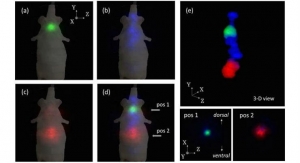
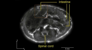
The G-SPECT also builds on the previously developed high resolution preclinical scanning technology of small animals.
“I am particularly impressed by the unprecedented spatial and temporal resolution I have seen as the first results. The G-SPECT may become a game changer in the medical imaging of various disorders of, for example, the brain, kidneys or joints," said professor Fred Verzijlbergen, head of the Department of Nuclear Medicine at the Erasmus Medical Center in Rotterdam. " In one go, we will get a wealth of detailed information from a single SPECT scan. The difference in resolution of the current SPECT technology is so big that it is difficult to say how great the revolution is the G-SPECT unleashes. Now, of course, thorough scientific testing on large groups of patients should be given high priority.”
Another advantage of the G-SPECT is that the technology is relatively inexpensive compared with positron emission tomography (PET) scanners that also can image radiolabeled tracers. In addition, G-SPECT allows clinicians to follow and simultaneously map different functions in a single scan, while in many cases only a low dose is needed.
The certification process for clinical use is in progress and expected to be finished early 2016. In 2011, MILabs introduced VECTor, an extremely user-friendly, fully integrated simultaneous SPECT and PET imaging technology with uniform and isotropic 0.75 mm resolution PET.
“The G-SPECT is the first SPECT scanner with a higher resolution than PET scanners. This may have a direct impact on millions of patients,” Beekman said.
MILabs was founded in 2006 as a spin-off from the University Medical Centre Utrecht, the Netherlands.