Rachel Klemovitch, Assistant Editor03.04.24
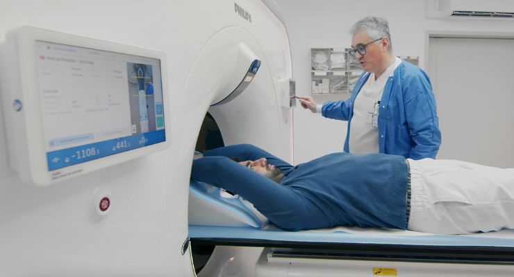
Royal Philips has released the Philips CT 5300 system with AI capabilities for diagnosis, interventional procedures, and screening. This X-ray CT system was released at #ECR2024.
The Philips CT 5300 system is designed to improve patient outcomes and increase diagnostic confidence by streamlining workflow efficiency and maximizing system up-time. The system can assist high-throughput, short-staffed radiology departments.
Recent research found a “CT first” strategy has improved patient care for those presenting chest pain with undiagnosed coronary artery disease. This strategy helped patients avoid more invasive procedures 1. However, there is a lack of available high-quality CT systems, for physicians who are qualified to read cardiac CT exams.
The CT 5300 system integrates virtual tools for real-time clinical and technical support. This helps overcome challenges related to increased patient loads, staff shortages, complex cases, and budget constraints.
“We listened to radiologists about the issues they face every day and what they wanted out of a next-generation CT system. We then combined the latest imaging and AI technologies to meet their needs and created a system that delivers next-level diagnostic confidence, empowers workflows, and ensures lifetime value,” said Frans Venker, Business Leader of CT at Philips. “We’ve leveraged AI in virtually every aspect of the CT 5300, freeing both CT technicians and radiologists from tedious, time-consuming tasks so they can spend more time focused on their patients.”
Philips’ system brings clinical expertise and high-end scanner functionality to an affordable and versatile system. The CT 5300 meets the requirements for advanced diagnostic imaging systems, cardiac patient guidelines, and addresses challenges in trauma care and interventional procedures.
The CT 5300 is currently installed in several European hospitals.
“CT 5300 offers CT imaging from head to toe, combined with high-end functionality such as coronary angiography, delivering an extremely broad spectrum of applications, helping us to better manage increased volumes of patients,” said Dr. Hilmar Kühl, Head of Radiology at St. Bernhard-Hospital Kamp-Lintfort, Germany. “With this latest system from Philips, we see an improvement in image quality with Precise Imaging, and for the first time, we can now visualize cardiac anatomy by using Precise Cardiac nearly artifact-free, which is very valuable to help improve cardiac care for our patients.”
The new system includes Philips Precise Image reconstruction software. It also introduces Nanopanel Precise, the industry’s first detector built from the ground up for AI-based reconstruction. CT 5300 uses a lower radiation dose to produce high-quality images.
When compared to conventional reconstruction, Precise Image archives 85% lower noise at an 80% lower dose and 60% better low-contrast detectability 2. When combined with Precise Cardiac motion compensation, Precise Image makes CT 5300 suitable for high-quality, motion-free, cardiac imaging in patients with high heart rates or heart-rate variability 3.
This system also features Philips CT Collaboration Live for remotely facilitated collaboration, training, and education.
Resources:
[1] Markus Scherer, MD, Atrium Health-Sanger Heart and Vascular Institute, Charlotte, NC. The study findings were presented February 1, 2024 at the American College of Cardiology (ACC) Cardiovascular Summit in Washington, DC.
[2] Lower image noise, improved low-contrast detectability, and/or dose reduction were tested using reference body protocols. All metrics were tested on phantoms. Low-contrast detectability tests were performed using 1.0 mm slices, and tested on the MITA CT IQ Phantom (CCT183, The Phantom Laboratory), using an auto tool “CHO” (Channelized Hotelling Observer). Data on file. Philips White Paper ‘Basic IMR testing, considerations and image quality trends’.
https://www.philips.co.uk/c-dam/b2bhc/gb/resource-catalog/landing/brightontender/ct-imr-white-paper-lr.pdf
[3] Precise Image for cardiac scans is 510(k) pending –not available for sale in the USA.
