Michael Barbella, Managing Editor04.01.21
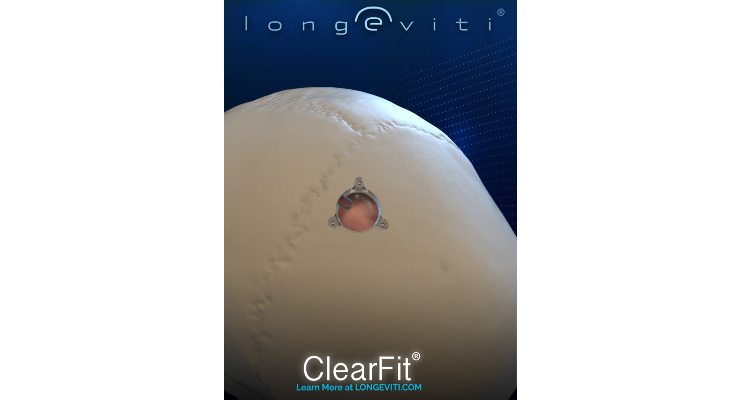
Longeviti Neuro Solutions has launched the Longeviti ClearFit Cover, a first-of-its-kind sonolucent cover used to replace excised cranial bone following a neurosurgical procedure. The product enables neurosurgeons to use ultrasound imaging to view neuroanatomy after brain surgery, allowing long-term monitoring of patients’ conditions.
“For decades we neurosurgeons have used burr holes and catheters in the treatment of hydrocephalus. We have often needed to use CT or MRI imaging to assess and optimize treatment. If we could use a similar burrhole, modified to fit an ultrasound-clear cover, to evaluate the size of the ventricles in our clinic, patient care would be greatly benefited. I think that burr hole ultrasounds have the potential of offering an easier, more convenient and less expensive way of optimizing our care, without radiation,” said Dr. Mark Luciano, M.D., Ph.D., FACS, director of Cerebral Fluid Center for Hydrocephalus and CSF Disorders at Johns Hopkins Hospital.
Patients with hydrocephalus and other neurological conditions such as subdural hematoma, epidural hematoma, brain tumors and traumatic brain injuries often require neurosurgery, and imaging for life. These neurological procedures may leave small holes in the skull to allow for insertion of a drain or catheter. Now, surgeons can use a Longeviti ClearFit Cover to repair the cranial void and serve as a neuroimaging diagnostic tool.
Longeviti ClearFit devices are implantable prosthetics used by neurosurgeons to correct and/or restore bony voids of a patient’s cranium. The ClearFit Cover is now part of a platform of ClearFit offerings from Longeviti, which includes off-the-shelf and customized patient-specific implants.
“Infants to seniors with hydrocephalus require repeat imaging through CT and MRI, not only for initial diagnosis of the condition, but in order to assess whether their treatment is working properly as well as post and pre-op for every surgical intervention. It is not uncommon for patients to require over 30 brain surgeries over the course of their lifetime,” said Diana Gray, president and CEO of the Hydrocephalus Association. “That translates into hundreds of imaging procedures, causing challenges including exposure to radiation, additional medical appointments, and increased medical costs. Technology that enables bedside imaging of the brain would reduce the burden on our families and could improve patient care.”
Hydrocephalus is the buildup of fluid in the ventricle deep within the brain. The excess fluid increases the size of the ventricles and puts pressure on the brain, causing headaches, dizziness and seizures, and patients with the condition can only be treated with brain surgery.
“In neurosurgery, imaging has been called the 'neurosurgeons' stethoscope.' Biometric data is key when making neurosurgical decisions on an operated patient. The keyhole created during time of craniotomy can be used as a sonolucent window, to reveal immediate post op changes at bedside. Using the ClearFit to fill in the “keyhole” we have the ability to ultrasound at the bedside and in the clinic,” said Dr. Netanel Ben-Shalom, M.D. of Israel’s Rabin Medical Center.
“We’ve learned from our neuro ICU nurses that 45.8 percent of patient transports are associated with adverse events,* not a surprise when you consider each 'transport' requires about five people and about seven connections per patient, often for imaging and diagnostics,” said Jesse Christopher, co-founder and CEO, Longeviti. “With ClearFit implants you can post-operatively image and upload video of the brain, virtually anywhere in the world, certainly bedside. We hope that this key feature can help reduce the frequency of patient transport, and ultimately help further reducing patient complications.
The ClearFit Cover is made of polymethyl-methacrylate (PMMA), a biocompatible material with over 40 years of proven clinical performance. It can also be used in conjunction with the Longeviti InvisiShunt, a neurosurgical implant that is placed in the cranium to restore the skull’s natural contour in patients undergoing complex brain surgeries.
* https://pubmed.ncbi.nlm.nih.gov/23587445/