Asociacion RUVID01.29.19
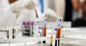
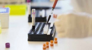
The method for diagnosing Alzheimer’s disease continues to be mostly clinical, which means it can’t be detected until the appearance of the first symptoms, or even later when the neuropathological damage is already severe. Thus, the search for new biomarkers that enable early detection of the disease in a non-invasive way becomes necessary.
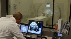
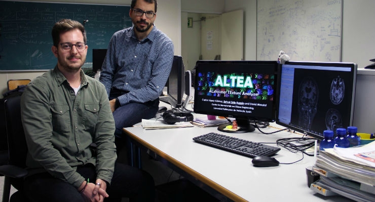
ALTEA takes on this challenge, a new tool created by researchers of Valencia’s Polytechnic University (UPV), belonging to the Centre of Biomaterials and Tissue Engineering (CBIT), in collaboration with Dr. Enrique Mollá, radiologist of the Hospital de La Ribera.
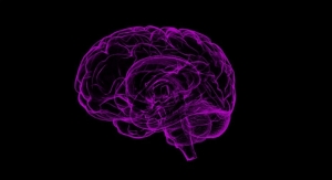
The tool, which is currently in a beta stage for research, would help detect Alzheimer’s disease in its most incipient stages. ALTEA makes it possible to visualize and segment magnetic resonance images, and based on these images, to extract and analyze different parameters linked to cerebral textures, transformed into biomarkers for Alzheimer’s disease (biomarkers are biological indicators that can be measured, and whose presence and intensity can be linked to the development of a disease).
“In light of the preliminary results obtained, we can say that both the analysis of 2D and 3D textures are very powerful tools that could complement and in great measure improve the diagnosis of Alzheimer’s disease. ALTEA could help with the early detection and to differentiate between the various stages of the disease,” highlighted David Moratal, a researcher for the Centre of Biomaterials and Tissue Engineering of the UPV.
How Does ALTEA Work?
What the software developed by the UPV researchers does, is a mathematic processing of the images, from which it extracts parameters that make it possible to quantify the homogeneity or heterogeneity of the hippocampal region. “With these parameters we can characterize and detect in what stage the disease is, and we can help reveal alterations that are invisible to the naked eye of clinical specialists,” added Rafael Ortíz, doctoral student at the UPV and one of the developers of the software together with UPV Biomedical Engineering Degree students Carlos López and Carolina Giménez.
Validation
To validate the new software, researchers analyzed magnetic resonance images belonging to three groups of people: patients with Alzheimer’s disease, patients with early minor cognitive impairment and individuals of a control group. The analyses were conducted on the hippocampal region (one of the areas most affected by brain atrophy in the first stages of the disease) using circular and spherical regions of interest. “Once ALTEA was developed, we conducted a transversal study with an extensive statistical analysis in order to verify the predictive capability of the potential biomarkers that were obtained, with very positive results,” explained David Moratal.
On its application in clinical practice, researchers said that “more texture parameters still have to be inserted, and the module that makes it possible to analyze combinations of parameters with automated learning techniques must be created, in order to create validated classifier models.”
ALTEA takes on this challenge, a new tool created by researchers of Valencia’s Polytechnic University (UPV), belonging to the Centre of Biomaterials and Tissue Engineering (CBIT), in collaboration with Dr. Enrique Mollá, radiologist of the Hospital de La Ribera.
The tool, which is currently in a beta stage for research, would help detect Alzheimer’s disease in its most incipient stages. ALTEA makes it possible to visualize and segment magnetic resonance images, and based on these images, to extract and analyze different parameters linked to cerebral textures, transformed into biomarkers for Alzheimer’s disease (biomarkers are biological indicators that can be measured, and whose presence and intensity can be linked to the development of a disease).
“In light of the preliminary results obtained, we can say that both the analysis of 2D and 3D textures are very powerful tools that could complement and in great measure improve the diagnosis of Alzheimer’s disease. ALTEA could help with the early detection and to differentiate between the various stages of the disease,” highlighted David Moratal, a researcher for the Centre of Biomaterials and Tissue Engineering of the UPV.
How Does ALTEA Work?
What the software developed by the UPV researchers does, is a mathematic processing of the images, from which it extracts parameters that make it possible to quantify the homogeneity or heterogeneity of the hippocampal region. “With these parameters we can characterize and detect in what stage the disease is, and we can help reveal alterations that are invisible to the naked eye of clinical specialists,” added Rafael Ortíz, doctoral student at the UPV and one of the developers of the software together with UPV Biomedical Engineering Degree students Carlos López and Carolina Giménez.
Validation
To validate the new software, researchers analyzed magnetic resonance images belonging to three groups of people: patients with Alzheimer’s disease, patients with early minor cognitive impairment and individuals of a control group. The analyses were conducted on the hippocampal region (one of the areas most affected by brain atrophy in the first stages of the disease) using circular and spherical regions of interest. “Once ALTEA was developed, we conducted a transversal study with an extensive statistical analysis in order to verify the predictive capability of the potential biomarkers that were obtained, with very positive results,” explained David Moratal.
On its application in clinical practice, researchers said that “more texture parameters still have to be inserted, and the module that makes it possible to analyze combinations of parameters with automated learning techniques must be created, in order to create validated classifier models.”