
6 Fraunhofer Research Demonstrations to See at Medica
By Fraunhofer-Gesellschaft | 11.12.18
Check out the latest research from Europe's largest application-oriented research organization.

Visit the Fraunhofer booths at the world's leading trade fair for the medical industry from 12th to 15th November 2018 in Düsseldorf to get a firsthand look at the following innovations poised to transform medical care:
1. Electrowetting (Hall 8a, Booth P13) and Telemedicine (Hall 10, Booth G05)
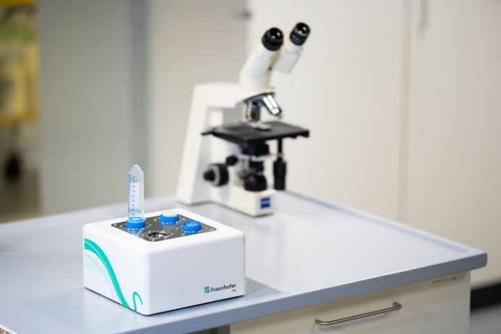
Researchers at Fraunhofer FIT have developed a system that now allows monitoring biochemical reactions at the single-molecule level from start to finish. It is based on the physical effect of Electrowetting-On-Dielectric (EWOD), widely used in microfluidics. The EWOD-BioPro system is capable of merging reagent droplets as small as 150nl with very high precision. This allows monitoring of the reaction process using a confocal microscope adapted at FIT, and to record high-resolution measurements.
"For the first time, we can observe the details of how the two droplets interact at the single-molecule level—thus allowing us to monitor the entire chain of events," said Lorenz Sparrenberg from the Fraunhofer Institute for Applied Information Technology FIT, who is leading the research project.
Biological interactions under conditions closely resembling those that occur naturally in the human body can this be studied. In pharmacology, for example, it is important to understand the effects of drug ingredients as precisely as possible and know when these effects occur. But the combination of EWOD and confocal microscopy may also be used to study the interaction between complementary DNA strands, antibodies and antigens, or enzyme-substrate reactions.
Conventional laboratory tests not only deliver less information but also require much higher quantities of the sample liquids, and take longer to perform. The typical duration of an enzymatic assay, for example, is around 15—20 minutes. The EWOD-BioPro system can present the results within 30 seconds. It may thus be a further step toward point-of-care diagnostics.
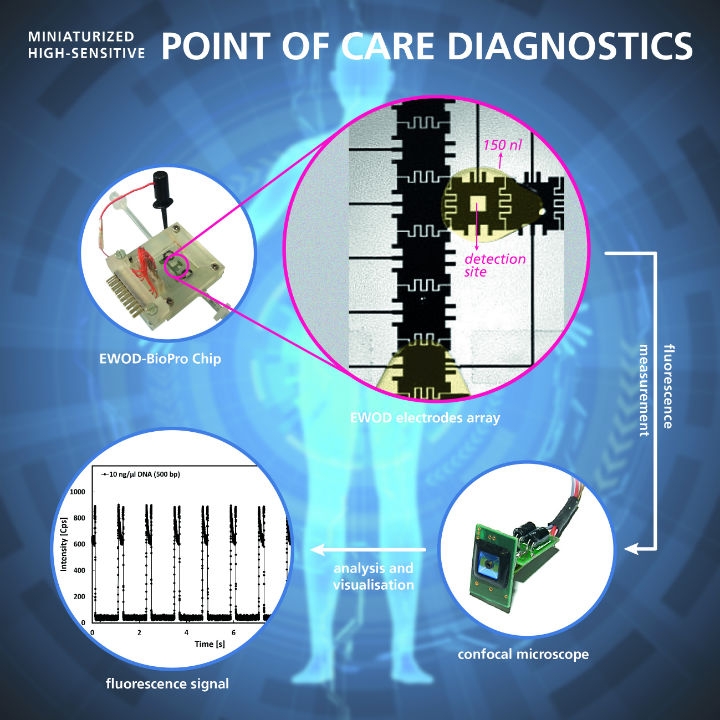
The EWOD-BioPro wetting-on-dielectric makes it easier to understand biological interactions. Image courtesy of Fraunhofer FIT.
The teliFIT platform features a modular, expandable architecture. It can accommodate a multitude of user groups with their specific access rights and can easily be tailored to a wide range of design requirements, contents, and functions. The platform's current functionalities may be grouped into five categories: Communication, Monitoring, Analysis, Smart Data Services, and Safety & Security. Individual functions can be activated or extended as required.
Smart devices can be used to capture and upload data to the teliFIT platform. Processing results are presented in visually attractive charts, but also offer relevant context. For example, teliFIT offers a nutrition diary that automatically evaluates the calories taken in against the user's individual basic requirement. Actual intake is also evaluated in relation to agreed nutritional goals.
Data security has been a major design criterion for the teliFIT platform. Personal data is routinely encrypted and stored in a separate database. The teliFIT platform is hosted in Germany. Its resilience is checked regularly through external penetration tests. There is a stable code base that includes high-quality software and architecture. Due to the use of state-of-the-art software technologies, the platform is easily scalable.
2. Artificial Intelligence for Optimal Patient Care (Hall 10, Stand G05)
Special software developed by the Fraunhofer Institute for Computer Graphics Research will soon assist health professionals in analysis of imaging data and also automatically generate a “virtual biopsy,” localizing and highlighting the tumor, displaying it three-dimensionally and analyzing the data. This will allow the software to derive over a hundred parameters from the CT images of a head and neck tumor. Initial results show that this approach not only analyzes CT images more quickly, it also provides information that could only be obtained through a surgical procedure and subsequent lab testing of the biopsied tissue. Aside from imaging organs and body regions, artificial intelligence is making it possible to automatically segment and analyze imaging data that is difficult to interpret.
Another interesting question doctors face is whether there are noticeable connections between the person being treated and other people. To answer this, health professionals combine the data of people with similar clinical pictures or other similarities, such as age or gender, into cohorts. The researchers at Fraunhofer IGD have developed software to assist doctors in forming suitable cohorts, examines them for significant connections, visualizes the attributes, and facilitates and accelerates the identification of clinically relevant hypotheses. What took several hours to do manually will take this automatic process mere seconds—valuable time gained for treating the patient. Incorporating artificial intelligence in the search for a hypothesis also ensures that a potentially critical factor is not overlooked.
Fraunhofer IGD’s visual solution combines all digitally available data into a graphic overview. Image courtesy of Fraunhofer IGD - everythingpossible/Fotolia.
Created by compiling all relevant parameters, the patient’s “digital twin” contributes to optimal care. Health@Hand, Fraunhofer IGD’s visual solution, combines all digitally available data, including the patient’s real-time vitals, into a graphic overview. As a digital control station, the system provides hospital staff with all relevant information with just a click and reprocesses it visually. This means necessary information is collected considerably more quickly. However, the control station is not content with just showing a single patient, rather it shows a live 3D model of the entire hospital ward, including inventory. Doctors and nurses can view the digital twin of the station on a PC or tablet and immediately know where, for example, a mobile X-ray machine is currently located. Key data for the entire ward can either be displayed all at once or viewed in detail for a single room or over a given period, for example. The goal is to simplify ward monitoring, detect disturbances immediately and thus be able to intervene in time.
To analyze an individual’s health information, the Health@Hand system links together crucial data from different clinical data systems to make possible entirely new conclusions. Trends in the patient’s health can be identified sooner and prognoses for the treatment process made more quickly. To maintain a patient’s health even at home, continuously recorded vitals can be supplied directly to the system. This would be sensible for, say, diabetes: The doctor immediately sees when levels exceed normal range and can take appropriate action. Even vitals and activity data from wearables, such as fitness armbands or smartwatches, can be included in the system, allowing it to act as a personal health assistant and make a valuable contribution to prevention.
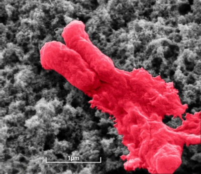
Antibiotic coating lyses bacterium (E. coli). Image courtesy of Fraunhofer IFAM.
3. No Chance for Bacteria on Implants (Hall 8a, Booth P13)
As part of the “Synergy-Boost” project, researchers at the Fraunhofer Institutes for Manufacturing Technology and Advanced Materials IFAM, for Molecular Biology and Applied Ecology IME, for Cell Therapy and Immunology IZI, and for Toxicology and Experimental Medicine ITEM have together developed a technique for preventing further hip joint infection. “We apply the antibiotic needed directly to the second implant—bringing it right to the spot where it’s needed,” said Kai Borcherding, a scientist at Fraunhofer IFAM. “We also did research on how antibiotics and silver ions work synergistically, significantly increasing the efficacy of the drug.” While both the antibiotic and the silver ions can eliminate pathogens, their combined effect is much stronger than the sum of their individual effects—they reinforce each other.
Silver ions have long been known to substantially enhance the effect of antibiotics, but research into the mechanism has been limited to isolated experiments. The optimal ratio of silver ions to antibiotic molecules is dependent not only on the drug used, but also on which microorganism is to be killed. The researchers at Fraunhofer IME initially performed large-scale screenings. They investigated 20 different antibiotics with various ratios of silver ions and on four key pathogens—a total of more than 9000 tests—to identify the most effective combinations.
If patients suffer from an infection and need a new hip joint or a new dental implant, the first thing the physician will do is to generate an antibiogram. Suitable sample material is taken, the microorganisms from it are cultivated and then analyzed to identify the appropriate antibiotic. This is a standard procedure. The physician then applies the appropriate antibiotic directly to the implant. “At Fraunhofer IFAM, we have already investigated how the antibiotics can be stored, and developed various coating methods to do this,” said Borcherding. The researchers have found a way to structure the surface so that it absorbs the antibiotic. In a vacuum, they apply the coating containing silver to the surface of the implant. Development of the coating process is now complete. Researchers at Fraunhofer IZI and Fraunhofer ITEM must now verify its efficacy. Researchers at Fraunhofer ITEM are also preparing the documentation required for approval of medical devices.
4. Individualized Therapy for Patients with Osteoporosis (Hall 10, Booth G05/H04)
Drug therapies are intended to inhibit bone depletion, but patients often do not respond to treatment. Studies also indicate a link between osteoporosis and cardiovascular diseases. Furthermore, the primary therapy for loss of bone mass is to increase calcium intake, the mineral is said to increase the stability of the bones. But raising the dosage of the mineral can lead to calcium deposits in veins, leading potentially to an increased risk of vascular occlusion and heart attack. The OsteoSys project therefore aims to investigate the interactions between cardiovascular diseases, inflammation, and bone metabolism, with a view to providing patients with personalized therapy and minimizing drug-related side effects.
Whether genetic or epigenetic (the influence of the environment on genes), the scientists take factors at the level of cells or organs into account to develop biomarkers and algorithms that predict adverse effects and enable patients to receive individualized treatment.
The role of the Fraunhofer FIT researchers in Sankt Augustin is to integrate data and create algorithms and models for the project. They are also establishing a sample management system and biobank management tools. “The software we develop to manage samples and laboratory procedures supports physicians in handling samples. Efficient data management supports our partners in their research work through the secure and traceable exchange of data, samples, and information,” said Carina Goretzky, a scientist at Fraunhofer FIT.
5. Interactive Shutter Eyeglasses to Replace Eyepatch Therapy (Hall 10, Booth G05/H04)
In a collaborative project, research and industry are aiming to achieve a decisive improvement in the often difficult therapy for young children with amblyopia (lazy eye). They hope that new interactive, context-dependent shutter eyeglasses with sensor-based feedback will increase treatment compliance for this complaint. This new technology covers up the patient’s good eye only when the situation is appropriate. During sport, for example, or other activities that demand good spatial vision, this function is deactivated so as to avoid any risk of accident.
Use and operation of the shutter eyeglasses are controlled by a multimodal sensor system integrated in the temples of the eyeglasses. Researchers at Fraunhofer Institute for Biomedical Engineering IBMT have developed this new technology along with a smartphone app to enable parents to monitor the treatment. All the data generated by this technology is compiled in a digital patient file system, which was also developed by the IBMT team. This web-based application is compliant with data-protection laws and can be accessed by the ophthalmologist in order to monitor, adjust and enhance treatment. For example, it tells the doctor if and when the patient has worn the eyeglasses—information that was never available with conventional treatment. “The data is uploaded to the app via Bluetooth and then securely archived in a cloud database,” explained Dr. Frank Ihmig, a scientist at Fraunhofer IBMT in St. Ingbert. “Our goal is to provide an individual, patient-based therapy.”
Functional model of electronic shutter eyeglasses. Image courtesy of Fraunhofer IBMT, Bernd Müller.
Data for real-time processing is generated by various sensors. These include temperature and skin-contact sensors to monitor the length of time and the position in which the eyeglasses have been worn. They also measure the occlusion phases, during which the LCD lenses are darkened. “This data is logged in an electronic memory device integrated in the eyeglass frames,” Ihmig explained. The eyeglass lenses are darkened by means of an electronically controlled shuttering system based on integrated liquid crystals. The frequency and duration of the occlusion therapy can be individually adjusted to each specific case. It is therefore more versatile than conventional eyepatch treatment. In this way, project partners hope to encourage young patients to wear the eyeglasses for the prescribed duration. The skin-contact sensors monitor whether the eyeglasses are being worn in the correct position and even provide the young wearer with child-friendly feedback. This can help increase acceptance for this form of therapy, too.
An acceleration sensor recognizes specific patterns of movement and can distinguish between different activities such as standing up, lying down, sitting down, walking, running, jumping, cycling and climbing stairs. “Our shutter eyeglasses are context-sensitive,” Ihmig explained. “This means that when the wearer is involved in energetic activities such as sport, for example, the mechanism for darkening the LCD lenses is deactivated, and they remain clear. This ensures full spatial vision and removes the risk of the wearer being involved in an accident.”
Initial tests with amblyopic children are scheduled for the second quarter of 2019. In addition, it is hoped that a validation study to be conducted toward the end of the project will confirm the medical benefits of the therapy.
Researchers have already developed a first functional model of the electronics for the new eyeglasses. The next step will be to make these smaller so they can be integrated in children’s eyeglass frames. In parallel, they are working to make the electronics more energy-efficient and thereby prolong battery life. The battery is recharged inductively, i.e., without a cable.
6. The Fast Way to Individual Treatment: Automated Tumor Diagnosis (Hall 10 Booth G05)
Fraunhofer researchers have developed an automated procedure for lymph node diagnosis as part of an inter-disciplinary project. The method provides important information on the stage of cancer: Cells from a primary tumor can make their way to other parts of the body through the lymph system and form metastases. Therefore, the presence of tumor cells in the lymph nodes means that the cancer has already spread and must be treated accordingly.
Until now, the lymph node tissue taken from patients has been solidified in paraffin in the laboratory, sliced into thin segments and examined under a microscope. “However, as pathologists are not able to examine all segments due to time constraints, some tumor cells may remain undiscovered,” explained Sebastian Schoening, group leader of the project group for Automation in Medicine and Biotechnology at Fraunhofer IPA.
The new method that the researchers from three Fraunhofer institutes have developed means that all tumor cells can be detected.
Lymph node diagnosis helps to determine whether a tumor has already spread across the body and started to form metastases. Image courtesy of Fraunhofer IPA.
“This is a radically different approach to previous lymph node diagnosis,” explained Schöning. Instead of cutting tissue, the samples are dissociated into individual cells. The grinding device required for this procedure, the so-called “Tissue Grinder,” was developed by the IPA team. The trick is that cells are being so carefully separated that they still function. In the next phase, tumor cells are colored, analyzed under the microscope, and counted. All of this is fully automated without a laboratory technician. The expertise for this system was contributed by the research team at the Fraunhofer Institute for Integrated Circuits IIS. The tumor cells are subsequently examined for genetic changes in order to select the optimal treatment for individual patients. This molecular test method was developed at the Fraunhofer Institute for Toxicology and Experimental Medicine ITEM in Regensburg. The software Merlin, developed by the IPA team, digitizes the entire procedure and makes it available in an electronic lab notebook covering all laboratory steps: from processing the samples to filing the findings report.
Automation not only makes the new LyDia HD diagnosis more precise but also quicker and more cost-effective than previous methods. Additionally, it immediately provides important information on the nature of the tumor cells for further treatment. This helps doctors to select suitable medication for the patient as part of a follow-up treatment plan.
1. Electrowetting (Hall 8a, Booth P13) and Telemedicine (Hall 10, Booth G05)
Researchers at Fraunhofer FIT have developed a system that now allows monitoring biochemical reactions at the single-molecule level from start to finish. It is based on the physical effect of Electrowetting-On-Dielectric (EWOD), widely used in microfluidics. The EWOD-BioPro system is capable of merging reagent droplets as small as 150nl with very high precision. This allows monitoring of the reaction process using a confocal microscope adapted at FIT, and to record high-resolution measurements.
"For the first time, we can observe the details of how the two droplets interact at the single-molecule level—thus allowing us to monitor the entire chain of events," said Lorenz Sparrenberg from the Fraunhofer Institute for Applied Information Technology FIT, who is leading the research project.
Biological interactions under conditions closely resembling those that occur naturally in the human body can this be studied. In pharmacology, for example, it is important to understand the effects of drug ingredients as precisely as possible and know when these effects occur. But the combination of EWOD and confocal microscopy may also be used to study the interaction between complementary DNA strands, antibodies and antigens, or enzyme-substrate reactions.
Conventional laboratory tests not only deliver less information but also require much higher quantities of the sample liquids, and take longer to perform. The typical duration of an enzymatic assay, for example, is around 15—20 minutes. The EWOD-BioPro system can present the results within 30 seconds. It may thus be a further step toward point-of-care diagnostics.
The EWOD-BioPro wetting-on-dielectric makes it easier to understand biological interactions. Image courtesy of Fraunhofer FIT.
The teliFIT platform features a modular, expandable architecture. It can accommodate a multitude of user groups with their specific access rights and can easily be tailored to a wide range of design requirements, contents, and functions. The platform's current functionalities may be grouped into five categories: Communication, Monitoring, Analysis, Smart Data Services, and Safety & Security. Individual functions can be activated or extended as required.
Smart devices can be used to capture and upload data to the teliFIT platform. Processing results are presented in visually attractive charts, but also offer relevant context. For example, teliFIT offers a nutrition diary that automatically evaluates the calories taken in against the user's individual basic requirement. Actual intake is also evaluated in relation to agreed nutritional goals.
Data security has been a major design criterion for the teliFIT platform. Personal data is routinely encrypted and stored in a separate database. The teliFIT platform is hosted in Germany. Its resilience is checked regularly through external penetration tests. There is a stable code base that includes high-quality software and architecture. Due to the use of state-of-the-art software technologies, the platform is easily scalable.
2. Artificial Intelligence for Optimal Patient Care (Hall 10, Stand G05)
Special software developed by the Fraunhofer Institute for Computer Graphics Research will soon assist health professionals in analysis of imaging data and also automatically generate a “virtual biopsy,” localizing and highlighting the tumor, displaying it three-dimensionally and analyzing the data. This will allow the software to derive over a hundred parameters from the CT images of a head and neck tumor. Initial results show that this approach not only analyzes CT images more quickly, it also provides information that could only be obtained through a surgical procedure and subsequent lab testing of the biopsied tissue. Aside from imaging organs and body regions, artificial intelligence is making it possible to automatically segment and analyze imaging data that is difficult to interpret.
Another interesting question doctors face is whether there are noticeable connections between the person being treated and other people. To answer this, health professionals combine the data of people with similar clinical pictures or other similarities, such as age or gender, into cohorts. The researchers at Fraunhofer IGD have developed software to assist doctors in forming suitable cohorts, examines them for significant connections, visualizes the attributes, and facilitates and accelerates the identification of clinically relevant hypotheses. What took several hours to do manually will take this automatic process mere seconds—valuable time gained for treating the patient. Incorporating artificial intelligence in the search for a hypothesis also ensures that a potentially critical factor is not overlooked.
Fraunhofer IGD’s visual solution combines all digitally available data into a graphic overview. Image courtesy of Fraunhofer IGD - everythingpossible/Fotolia.
Created by compiling all relevant parameters, the patient’s “digital twin” contributes to optimal care. Health@Hand, Fraunhofer IGD’s visual solution, combines all digitally available data, including the patient’s real-time vitals, into a graphic overview. As a digital control station, the system provides hospital staff with all relevant information with just a click and reprocesses it visually. This means necessary information is collected considerably more quickly. However, the control station is not content with just showing a single patient, rather it shows a live 3D model of the entire hospital ward, including inventory. Doctors and nurses can view the digital twin of the station on a PC or tablet and immediately know where, for example, a mobile X-ray machine is currently located. Key data for the entire ward can either be displayed all at once or viewed in detail for a single room or over a given period, for example. The goal is to simplify ward monitoring, detect disturbances immediately and thus be able to intervene in time.
To analyze an individual’s health information, the Health@Hand system links together crucial data from different clinical data systems to make possible entirely new conclusions. Trends in the patient’s health can be identified sooner and prognoses for the treatment process made more quickly. To maintain a patient’s health even at home, continuously recorded vitals can be supplied directly to the system. This would be sensible for, say, diabetes: The doctor immediately sees when levels exceed normal range and can take appropriate action. Even vitals and activity data from wearables, such as fitness armbands or smartwatches, can be included in the system, allowing it to act as a personal health assistant and make a valuable contribution to prevention.
Antibiotic coating lyses bacterium (E. coli). Image courtesy of Fraunhofer IFAM.
As part of the “Synergy-Boost” project, researchers at the Fraunhofer Institutes for Manufacturing Technology and Advanced Materials IFAM, for Molecular Biology and Applied Ecology IME, for Cell Therapy and Immunology IZI, and for Toxicology and Experimental Medicine ITEM have together developed a technique for preventing further hip joint infection. “We apply the antibiotic needed directly to the second implant—bringing it right to the spot where it’s needed,” said Kai Borcherding, a scientist at Fraunhofer IFAM. “We also did research on how antibiotics and silver ions work synergistically, significantly increasing the efficacy of the drug.” While both the antibiotic and the silver ions can eliminate pathogens, their combined effect is much stronger than the sum of their individual effects—they reinforce each other.
Silver ions have long been known to substantially enhance the effect of antibiotics, but research into the mechanism has been limited to isolated experiments. The optimal ratio of silver ions to antibiotic molecules is dependent not only on the drug used, but also on which microorganism is to be killed. The researchers at Fraunhofer IME initially performed large-scale screenings. They investigated 20 different antibiotics with various ratios of silver ions and on four key pathogens—a total of more than 9000 tests—to identify the most effective combinations.
If patients suffer from an infection and need a new hip joint or a new dental implant, the first thing the physician will do is to generate an antibiogram. Suitable sample material is taken, the microorganisms from it are cultivated and then analyzed to identify the appropriate antibiotic. This is a standard procedure. The physician then applies the appropriate antibiotic directly to the implant. “At Fraunhofer IFAM, we have already investigated how the antibiotics can be stored, and developed various coating methods to do this,” said Borcherding. The researchers have found a way to structure the surface so that it absorbs the antibiotic. In a vacuum, they apply the coating containing silver to the surface of the implant. Development of the coating process is now complete. Researchers at Fraunhofer IZI and Fraunhofer ITEM must now verify its efficacy. Researchers at Fraunhofer ITEM are also preparing the documentation required for approval of medical devices.
4. Individualized Therapy for Patients with Osteoporosis (Hall 10, Booth G05/H04)
Drug therapies are intended to inhibit bone depletion, but patients often do not respond to treatment. Studies also indicate a link between osteoporosis and cardiovascular diseases. Furthermore, the primary therapy for loss of bone mass is to increase calcium intake, the mineral is said to increase the stability of the bones. But raising the dosage of the mineral can lead to calcium deposits in veins, leading potentially to an increased risk of vascular occlusion and heart attack. The OsteoSys project therefore aims to investigate the interactions between cardiovascular diseases, inflammation, and bone metabolism, with a view to providing patients with personalized therapy and minimizing drug-related side effects.
Whether genetic or epigenetic (the influence of the environment on genes), the scientists take factors at the level of cells or organs into account to develop biomarkers and algorithms that predict adverse effects and enable patients to receive individualized treatment.
The role of the Fraunhofer FIT researchers in Sankt Augustin is to integrate data and create algorithms and models for the project. They are also establishing a sample management system and biobank management tools. “The software we develop to manage samples and laboratory procedures supports physicians in handling samples. Efficient data management supports our partners in their research work through the secure and traceable exchange of data, samples, and information,” said Carina Goretzky, a scientist at Fraunhofer FIT.
5. Interactive Shutter Eyeglasses to Replace Eyepatch Therapy (Hall 10, Booth G05/H04)
In a collaborative project, research and industry are aiming to achieve a decisive improvement in the often difficult therapy for young children with amblyopia (lazy eye). They hope that new interactive, context-dependent shutter eyeglasses with sensor-based feedback will increase treatment compliance for this complaint. This new technology covers up the patient’s good eye only when the situation is appropriate. During sport, for example, or other activities that demand good spatial vision, this function is deactivated so as to avoid any risk of accident.
Use and operation of the shutter eyeglasses are controlled by a multimodal sensor system integrated in the temples of the eyeglasses. Researchers at Fraunhofer Institute for Biomedical Engineering IBMT have developed this new technology along with a smartphone app to enable parents to monitor the treatment. All the data generated by this technology is compiled in a digital patient file system, which was also developed by the IBMT team. This web-based application is compliant with data-protection laws and can be accessed by the ophthalmologist in order to monitor, adjust and enhance treatment. For example, it tells the doctor if and when the patient has worn the eyeglasses—information that was never available with conventional treatment. “The data is uploaded to the app via Bluetooth and then securely archived in a cloud database,” explained Dr. Frank Ihmig, a scientist at Fraunhofer IBMT in St. Ingbert. “Our goal is to provide an individual, patient-based therapy.”
Functional model of electronic shutter eyeglasses. Image courtesy of Fraunhofer IBMT, Bernd Müller.
Data for real-time processing is generated by various sensors. These include temperature and skin-contact sensors to monitor the length of time and the position in which the eyeglasses have been worn. They also measure the occlusion phases, during which the LCD lenses are darkened. “This data is logged in an electronic memory device integrated in the eyeglass frames,” Ihmig explained. The eyeglass lenses are darkened by means of an electronically controlled shuttering system based on integrated liquid crystals. The frequency and duration of the occlusion therapy can be individually adjusted to each specific case. It is therefore more versatile than conventional eyepatch treatment. In this way, project partners hope to encourage young patients to wear the eyeglasses for the prescribed duration. The skin-contact sensors monitor whether the eyeglasses are being worn in the correct position and even provide the young wearer with child-friendly feedback. This can help increase acceptance for this form of therapy, too.
An acceleration sensor recognizes specific patterns of movement and can distinguish between different activities such as standing up, lying down, sitting down, walking, running, jumping, cycling and climbing stairs. “Our shutter eyeglasses are context-sensitive,” Ihmig explained. “This means that when the wearer is involved in energetic activities such as sport, for example, the mechanism for darkening the LCD lenses is deactivated, and they remain clear. This ensures full spatial vision and removes the risk of the wearer being involved in an accident.”
Initial tests with amblyopic children are scheduled for the second quarter of 2019. In addition, it is hoped that a validation study to be conducted toward the end of the project will confirm the medical benefits of the therapy.
Researchers have already developed a first functional model of the electronics for the new eyeglasses. The next step will be to make these smaller so they can be integrated in children’s eyeglass frames. In parallel, they are working to make the electronics more energy-efficient and thereby prolong battery life. The battery is recharged inductively, i.e., without a cable.
6. The Fast Way to Individual Treatment: Automated Tumor Diagnosis (Hall 10 Booth G05)
Fraunhofer researchers have developed an automated procedure for lymph node diagnosis as part of an inter-disciplinary project. The method provides important information on the stage of cancer: Cells from a primary tumor can make their way to other parts of the body through the lymph system and form metastases. Therefore, the presence of tumor cells in the lymph nodes means that the cancer has already spread and must be treated accordingly.
Until now, the lymph node tissue taken from patients has been solidified in paraffin in the laboratory, sliced into thin segments and examined under a microscope. “However, as pathologists are not able to examine all segments due to time constraints, some tumor cells may remain undiscovered,” explained Sebastian Schoening, group leader of the project group for Automation in Medicine and Biotechnology at Fraunhofer IPA.
The new method that the researchers from three Fraunhofer institutes have developed means that all tumor cells can be detected.
Lymph node diagnosis helps to determine whether a tumor has already spread across the body and started to form metastases. Image courtesy of Fraunhofer IPA.
“This is a radically different approach to previous lymph node diagnosis,” explained Schöning. Instead of cutting tissue, the samples are dissociated into individual cells. The grinding device required for this procedure, the so-called “Tissue Grinder,” was developed by the IPA team. The trick is that cells are being so carefully separated that they still function. In the next phase, tumor cells are colored, analyzed under the microscope, and counted. All of this is fully automated without a laboratory technician. The expertise for this system was contributed by the research team at the Fraunhofer Institute for Integrated Circuits IIS. The tumor cells are subsequently examined for genetic changes in order to select the optimal treatment for individual patients. This molecular test method was developed at the Fraunhofer Institute for Toxicology and Experimental Medicine ITEM in Regensburg. The software Merlin, developed by the IPA team, digitizes the entire procedure and makes it available in an electronic lab notebook covering all laboratory steps: from processing the samples to filing the findings report.
Automation not only makes the new LyDia HD diagnosis more precise but also quicker and more cost-effective than previous methods. Additionally, it immediately provides important information on the nature of the tumor cells for further treatment. This helps doctors to select suitable medication for the patient as part of a follow-up treatment plan.