Dawn A. Lissy, President, Empirical06.06.23
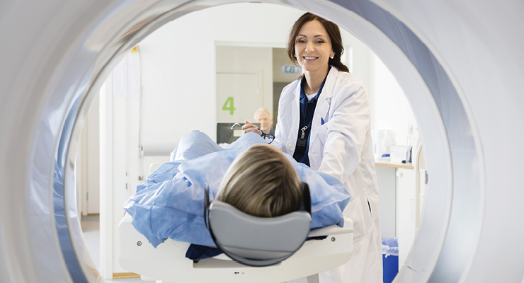
I did my best to lie perfectly still as the computed tomography (CT) scanner whirred around me, shooting narrow x-ray beams through my abdomen. The tomographic images would give us 3D views of exactly what was going on in my spine. From there, we could come up with a treatment plan for how to deal with my increasing discomfort.
Thanks to the CT scan, I know exactly where the breakdown was occurring and what we could do to fix it. It’s essentially the same process that is being increasingly applied to medical devices, particularly the smaller lots and custom parts being additively manufactured.
“It’s an endless loop. It becomes, ‘OK, I can’t use this part any more once I cut it,’” said Ed Rajchel, metrology inspection manager for Applied Technical Services. “Especially if it’s a custom part. You could take a metallurgical section, but you can’t use the part any more. The only way to look at the inside accurately is via CT scan.”
As additive manufacturing in healthcare continues to grow (2021’s market size was $1.6 billion), it’s changing the way we manufacture life-saving and life-enhancing devices. When medtech is being produced a single piece at a time, the processes by which its safety is tested have to adapt. When you’re producing a custom device, it doesn’t make sense to cut into it or destroy it to assess its limits when a microCT scan offers a clear glimpse of what’s inside without breaking down or into the device.
“If your doctor wanted to look in your brain, they could cut into you, which may not be ideal,” Rajchel said. “We can do the same thing with industrial parts. It may not always be ideal to cut it open. We can scan a part and look at the inside of it [to see] porosity, cracks, lack of fusion in the material.”
With microCT scanning, device developers can measure a product against its initial CAD model, he said.
“Given sometimes orthopedic implants can have complex geometries, it makes sense to scan,” Rajchel said. “Features such as reverse threads or embedded channels render it nearly impossible to physically touch and inspect these parts without destructive means.”
CT scanning technology started out by building layers of fan beam images and transitioned to the cone beam systems we see today. Scanners of yesteryear had significantly lower resolution and took exponentially longer.
The complexity and capability of microCT scanning is growing and adapting to meet the demands of product developers. Current technology enables medical device developers to assess if an additively manufactured implant is sterile and whether residual powder from manufacturing will affect the function of the device. With microCT scanning, we can pinpoint a wide variety of issues unique to standards and concerns for safe implantation in the human body.
“The beauty of 3D printing is, you can have a lot more complex internal geometries given the way we can print parts,” Rajchel said. “The only way to inspect the internal features of a part is by some kind of X-ray technology outside of cutting it open. We no longer have to do that because the X-ray technology allows us to see in inside the parts accurately. It’s becoming more and more accurate.”
It's a process that’s easily outsourced, which Rajchel said is a much more practical option than trying to set up in-house microCT scanning equipment.
“It’s a no-brainer. You spend a million dollars-plus [for machinery],” he said. “That’s for the machine alone—then you also have to pay for employees, software, calibration, the footprint. [For manufacturers], any space you’re not manufacturing is lost revenue.”
The cost of a microCT scan is slightly more expensive than destructive testing, Rajchel said. But you also end up with a part you can still use.
“I think a lot of it is, what is the actual part worth to you?” Rajchel said. “It’s all relative to the problem and the cost benefit of what the overall problem is. It might be nominally more than taking a metallurgical cross section. But not much.”
That’s why Rajchel is seeing anecdotal increases in medical device developers who are adding microCT scanning to their battery of tests as they push their products to market.
“As some of the OEM process centers are starting to incorporate it, I think it’s more widely accepted. It’s the only solution for the market,” Rajchel said. “As [medical devices] become more and more complex, you need more and more complex inspection methods.”
From the regulatory side of medical device development, there’s no specific indication for microCT scanning. But a scan can add value to—or possibly, eventually replace—the need for tension bar testing and in some cases, static and fatigue testing depending on the device and its application. It’s a tool that can directly impact and improve quality systems and parts validation.
But there’s also a challenge unique to the medical device community: We don’t have a standard accepted practice for lot release testing on additive parts. The intention is to ensure additively manufactured components will perform as designed and intended, but there’s no universal standard to determine that. Some manufacturers test every single plate. Some developers perform one test per day of manufacturing. Others will test one product per lot of 500.
MicroCT scanning can provide information and analysis that could address tensile, mechanical, static, and dynamic testing of a finished product. These scans can also deliver information critical for the validation and point of failure of additively manufactured devices.
“You can do many different tests within one test,” Rajchel said. “We can look at the internal microstructure of the part and do the dimensional inspection all in the same scan. There are some parallel processes that can be lumped into one test. It can definitely help people bring [medical devices] to market faster.”
As I travel the country for client visits and to speak at events, I’m often asked about what’s new or exciting in the world of testing. My answer this year is the potential for microCT scanning to revolutionize the way we assess the safety and efficacy of medical devices, especially the growing number coming from additive manufacturing. We now have a testing technology that can quickly identify issues in a device—without destroying it—before it ends up in the human body. It’s a win for device developers, patients, and our industry as a whole.
Dawn Lissy is a biomedical engineer, entrepreneur, and innovator. Since 1998, Empirical Technologies Corp. has operated under Lissy’s direction. Empirical offers the full range of regulatory and quality systems consulting, testing, small batch and prototype manufacturing, and validations services to bring a medical device to market. Empirical is very active within standards development organization ASTM International and has one of the widest scopes of test methods of any accredited independent lab in the United States. Because Lissy was a member of the U.S. Food and Drug Administration’s Entrepreneur-in-Residence program, she has first-hand, in-depth knowledge of the regulatory landscape. Lissy holds an inventor patent for the Stackable Cage System for corpectomy and vertebrectomy. Her M.S. in biomedical engineering is from The University of Akron, Ohio.
