Videos
3D Printers Produce Models that Enhance a Surgeon's Capabilities
3D Printers Produce Models that Enhance a Surgeon's Capabilities
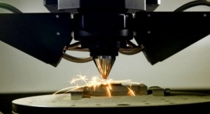
A specialized but fairly affordable printer produces three-dimensional models of an individual’s skull or body part.
By UC Davis Health03.04.19
Imagine 1,000 puzzle pieces without any picture of what it’s ultimately supposed to look like. With few, if any, reference points, the challenge of fitting them together would be daunting.
That’s what surgeons often confront when a patient suffering from a traumatic injury or condition has a portion of their body that is dramatically damaged or changed. The “puzzle” can be exponentially harder when the injuries involve a person’s face or skull—areas of the human anatomy that are complex, difficult to surgically navigate, and often require both functional and near-perfect cosmetic repair.
Now, thanks to high-tech equipment that is sometimes not much bigger than a home printer, UC Davis Health physicians are enhancing their capabilities and mapping out surgeries in ways that benefit patients and surgical outcomes.
Game-Changing Technology
“3D printing, which for us means manufacturing that’s accurate, affordable and on-site, can be a game changer in health care,” said David Lubarsky, vice chancellor for Human Health Sciences and CEO of UC Davis Health, who is very encouraged by the university’s newest technology initiatives and promising results.
The new device is a specialized but fairly affordable printer that produces three-dimensional models of an individual’s skull or body part. The 3D models enable a surgeon to visualize, practice and then perform the reconstructive surgery while saving time and increasing precision.
“Facial reconstructive surgery involves intricate anatomy within an extremely narrow operative field in which to maneuver our instruments,” said E. Bradley Strong, a professor of otolaryngology who specializes in facial reconstructive surgery. “Being able to print out a high-resolution 3D model of the injury, allows us to do detailed preoperative planning and preparation that is more efficient and accurate. We can also use these patient-specific models in the operating room to improve the accuracy of implant placement.”
Creating Models that Save Time in the Operating Room
The 3D printer used by Strong and his colleagues for the past year is about the size of a mini-refrigerator and costs approximately $4,000. It uses the imaging data from a patient’s computed tomography (CT) scans to provide the modeling output information. Like an inkjet printer, the 3D version spits out layer upon layer of material over a period of hours, sometimes taking nearly a day to complete, depending on the complexity of the model. The finished replica can save time during surgery, which means less time on the operating table for a patient and potentially a better outcome.
By creating a 3D model prior to surgery, Strong is able to bend and customize generic surgical plates into patient-specific shapes that fit perfectly for each individual patient.
Technology That Is Patient-Specific
For Joseph Michael, 3D printing was a key to regaining normal vision in his right eye, which had been severely damaged during a brutal assault at his home in 2013. Michael’s orbital socket was damaged when he was struck in the face by several assailants. His eye socket noticeably dropped down, causing double vision.
“I couldn’t drive before my surgery with Dr. Strong,” said the retired Grass Valley resident, whose injuries required about seven-and-a-half hours of surgical repair.
Strong told his patient about the 3D printing process, which involved using the CT scan of Michael’s left eye and making a virtual 3D model on the computer. The virtual model of his left eye was then mirrored to the right side to generate a precise template that could be used to reconstruct the damaged right eye. The right eye model was then printed and sterilized for surgery. Strong then used the model to precisely bend the hardware intraoperatively to repair Michael’s orbit and cheekbone.
“Overall, my friends up here [in Grass Valley] and even my eye doctor can’t believe the difference,” said Michael, regarding his appearance and the restored balance in the level of his eyes. “Dr. Strong gave me my eyesight back.”
A New Approach to Surgical Planning
The gold-standard for training and planning in difficult surgery cases has traditionally been through the use of cadaver models. While cadavers may have excellent anatomic and physical features, they aren’t always readily accessible. More importantly, they lack patient-specific details that are very helpful for surgeons. Printing a three-dimensional replica based on the imaging information from an individual patient is like working with a mirror image of that person. It can be highly detailed and fairly exact.
“With or without a 3D model, Mr. Michael’s surgical incisions would have been very similar,” Strong noted. “But the accuracy of pre-surgical planning and the precision of implant bending and placement were greatly improved. It saves time and enables me to be more efficient and make critical decisions before entering the operating room.”
A Model for Training and Education
Beyond surgery, 3D printers that can replicate the complexity and range of patient pathologies are excellent tools for medical research, education, and training. Surgery residents at UC Davis Medical Center are getting more and more opportunities to use printouts of specific body parts, enabling them to better plan, practice and achieve an optimal surgical result. Strong and colleagues that include Toby Steele have published studies showing the benefits of the high-resolution, patient-specific printed models.
Collaboration Is the Best Medicine
Three-dimensional printing applications and research work are not limited to the School of Medicine. Similar initiatives are underway at UC Davis School of Veterinary Medicine, where the work is complemented by the university’s Department of Biomedical Engineering expertise. The unique combination of disciplines provides opportunities to advance health that few other universities in the world are able to pursue. Veterinary surgeons and radiologists, for example, are working to validate the geometric accuracy of 3D replicas, which are also being used in companion animal and other veterinary cases since the modeling—like that for human patients—is all based on CT scanning data.
“There is great value in working together because veterinary medicine is a bit more 'nimble' on the development and application side, but the patient needs are very similar,” said Denis Marcellin-Little, professor of small animal orthopedic surgery who, along with colleague Boaz Arzi, has been discussing the use of 3D technologies and collaborative opportunities with colleagues in the School of Medicine.
The schools of medicine and veterinary medicine are also looking to the expertise in Biomedical Engineering, which has a production and manufacturing lab filled with an array of 3D printers, from simple and inexpensive models to complex and costly ones. The machines can process a variety of material types for creating specialized models and their working prototypes that offer a glimpse of the future when three-dimensional printers might produce biocompatible tissue and bone printouts.
Translating Engineering Advances to Medicine Means TEAM Work
Currently, the department’s Translating Engineering Advances to Medicine (TEAM) Lab is printing the 3D body parts used in veterinary surgeries. It has also worked with oncologists and physicists in the UC Davis Department of Radiation Oncology to fabricate what are called boluses—custom-printed sheets of plastic “skin” that conform precisely to a patient’s cancer treatment area—that help increase the accuracy of a radiation dose while also reducing it in tissues beyond the depth of a patient’s tumor.
The TEAM lab expertise, which is already helping clinicians bring their research to life to advance clinical care for humans and animals, demonstrates the promise of 3D technology and its potential for an entire UC Davis program to be built around it.
“By enabling intricate anatomy to be translated from computerized data into life-like replicas that clinicians can actually hold, analyze and work with means that challenging medical puzzles will become that much easier to solve,” Lubarsky added.
Related Searches: