Sam Brusco, Associate Editor03.29.22
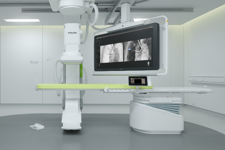
Philips has installed new Philips Lung Suite 3D real-time lung cancer imaging solutions at partner sites in London, Brussels, Genk, Belgium; Haifa, Israel; and one is soon to be installed in Rouen, France. The Lung Suite is used in combination with the company’s Image Guided Therapy System – Azurion.
Lung Suite addressed the need for earlier diagnosis and minimally-invasive treatment with a platform that allows doctors to perform biopsy, ablation, lesion marking, and/or thoracic surgery in the same room.
“Philips Lung Suite has been shown to increase the accuracy of lung cancer biopsy procedures, improving results for patients and offering the potential to immediately treat early-stage lung cancer patients,” Karim Boussebaa, GM, Image Guided Therapy Systems at Philips told the press. “These new partners add to our rapidly expanding global ecosystem of clinical partners who are pushing innovation forward, with of the goal of offering patients diagnosis and minimally-invasive treatment in a single procedure, improving patient outcomes and their quality of life.”
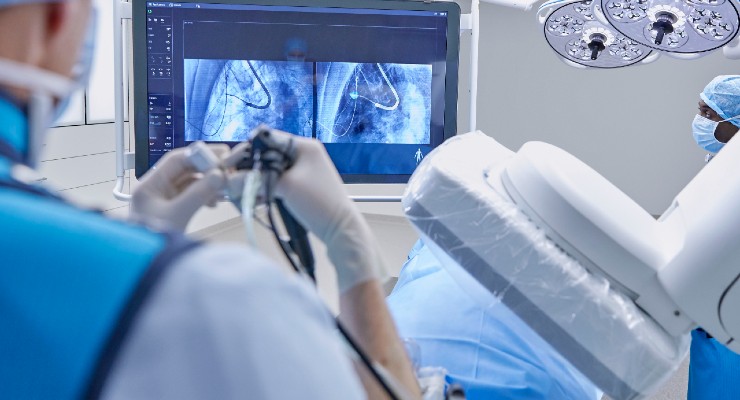
Lung Suite offers advanced 3D real-time imaging with augmented fluoroscopy on inage guided therapy systems, combined with dedicated software. Cone beam CT imaging generates a CT-like image in about five seconds, rotating around the patient to produce a high-resolution 3D view of the target lesion and other anatomical structures.
This lets the clinician performing a biopsy to be continually guided to advance a catheter toward the lesion through a bronchoscope. Once done, its position can be confirmed using the same imaging modality. The biopsy can then be taken.
“In the fast-growing world of intraoperative imaging, cone-beam CT remains the gold standard for augmented fluoroscopy and lesion confirmation. Using Lung Suite, no nodule can hide, regardless of anatomical position or radiologic characteristic, making it a valuable tool for both diagnosis and future ablation procedures,” said Dr. Amir Abramovich, MD, Director of Interventional Pulmonology at the Carmel Medical Center in Haifa, Israel.
“Cone-beam CT is the critical step towards targeting sub-20 mm nodules and an essential tool for the transition towards bronchoscopic microwave ablation of peripheral lung lesions,” said Professor Shah Pallav, MD, consultant respiratory physician at Royal Brompton Hospital in London, UK.
“The advanced cone beam CT imaging combined with augmented fluoroscopy of Philips Lung Suite gives us the confidence to safely reach and biopsy difficult-to-access peripheral lung nodules,” said Maarten Criel, MD, Pulmonologist at ZOL Genk Medical Center, Belgium.
Lung Suite addressed the need for earlier diagnosis and minimally-invasive treatment with a platform that allows doctors to perform biopsy, ablation, lesion marking, and/or thoracic surgery in the same room.
“Philips Lung Suite has been shown to increase the accuracy of lung cancer biopsy procedures, improving results for patients and offering the potential to immediately treat early-stage lung cancer patients,” Karim Boussebaa, GM, Image Guided Therapy Systems at Philips told the press. “These new partners add to our rapidly expanding global ecosystem of clinical partners who are pushing innovation forward, with of the goal of offering patients diagnosis and minimally-invasive treatment in a single procedure, improving patient outcomes and their quality of life.”
Lung Suite offers advanced 3D real-time imaging with augmented fluoroscopy on inage guided therapy systems, combined with dedicated software. Cone beam CT imaging generates a CT-like image in about five seconds, rotating around the patient to produce a high-resolution 3D view of the target lesion and other anatomical structures.
This lets the clinician performing a biopsy to be continually guided to advance a catheter toward the lesion through a bronchoscope. Once done, its position can be confirmed using the same imaging modality. The biopsy can then be taken.
“In the fast-growing world of intraoperative imaging, cone-beam CT remains the gold standard for augmented fluoroscopy and lesion confirmation. Using Lung Suite, no nodule can hide, regardless of anatomical position or radiologic characteristic, making it a valuable tool for both diagnosis and future ablation procedures,” said Dr. Amir Abramovich, MD, Director of Interventional Pulmonology at the Carmel Medical Center in Haifa, Israel.
“Cone-beam CT is the critical step towards targeting sub-20 mm nodules and an essential tool for the transition towards bronchoscopic microwave ablation of peripheral lung lesions,” said Professor Shah Pallav, MD, consultant respiratory physician at Royal Brompton Hospital in London, UK.
“The advanced cone beam CT imaging combined with augmented fluoroscopy of Philips Lung Suite gives us the confidence to safely reach and biopsy difficult-to-access peripheral lung nodules,” said Maarten Criel, MD, Pulmonologist at ZOL Genk Medical Center, Belgium.
