
Sam Brusco, Associate Editor12.16.21
Kheiron Medical Technologies, a company applying machine learning to cancer diagnostics, began collaborating with Stanford University researchers to build functional, proof-of-concept deep learning models for clinical problem-solving, beginning with non-Hodgkin’s lymphoma.
Kheiron is a pioneer of AI solutions to help radiologists spot breast cancer earlier. Its flagship Mia (Mammography Intelligent Assesment) AI platform for breast screening helps radiologists make the critical breast-screening decision of whether or not to recall.
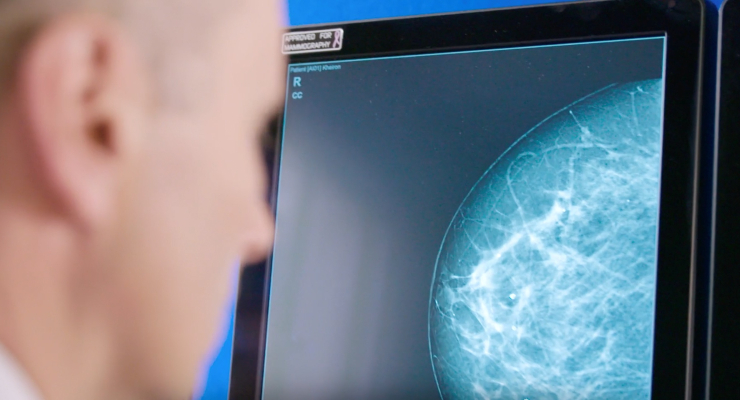
Image courtesy of Kheiron Medical Technologies.
“The Kaplan Project”—named in honor of former Stanford radiation oncology leader Dr. Henry Kaplan who developed some of the earliest lymphoma treatments—will Apply Kheiron’s deep learning technology to FEG-PET/CT scans of lymphoma patients to improve radiologist efficiency and accuracy.
Both organizations aim to utilize interdisciplinary expertise to solve critical clinical problems using AI.
“Projects like this one are so exciting because they capitalize on collaborations not only between clinicians and data scientists, but also between academics and industry,” AIMI Center director Dr. Curt Langlotz told the press.
Staging lymphoma quantifies the disease’s extent, guides therapy decisions, and gives a baseline before treatment. Staging and assessing post-treatment response on PET/CT image is also manual and time-consuming for oncological radiologists.
“This groundbreaking project marks a new chapter in the application of AI to transform cancer diagnostics across the entire patient pathway,” said Kheiron CEO Dr. Peter Kecskemethy. “Uniting new deep learning technologies with the clinical expertise of academic research institutions like Stanford will lead to the development of a completely new category of AI diagnostics and ultimately improve patient outcomes.”
“Our project aims to improve a time-consuming and mostly qualitative process, the longitudinal assessment of whole-body FDG-PET/CT scans, using deep learning to augment imaging specialists,” said Dr. Guido A. Davidzon, Clinical Associate Professor at Stanford University. “The overarching goal is to reduce the time needed to evaluate a PET scan, and by improving our throughput, ultimately increase patient access to a well-established noninvasive diagnostic imaging tool used by oncologists in the care of cancer.”
Kheiron is a pioneer of AI solutions to help radiologists spot breast cancer earlier. Its flagship Mia (Mammography Intelligent Assesment) AI platform for breast screening helps radiologists make the critical breast-screening decision of whether or not to recall.
Image courtesy of Kheiron Medical Technologies.
“The Kaplan Project”—named in honor of former Stanford radiation oncology leader Dr. Henry Kaplan who developed some of the earliest lymphoma treatments—will Apply Kheiron’s deep learning technology to FEG-PET/CT scans of lymphoma patients to improve radiologist efficiency and accuracy.
Both organizations aim to utilize interdisciplinary expertise to solve critical clinical problems using AI.
“Projects like this one are so exciting because they capitalize on collaborations not only between clinicians and data scientists, but also between academics and industry,” AIMI Center director Dr. Curt Langlotz told the press.
Staging lymphoma quantifies the disease’s extent, guides therapy decisions, and gives a baseline before treatment. Staging and assessing post-treatment response on PET/CT image is also manual and time-consuming for oncological radiologists.
“This groundbreaking project marks a new chapter in the application of AI to transform cancer diagnostics across the entire patient pathway,” said Kheiron CEO Dr. Peter Kecskemethy. “Uniting new deep learning technologies with the clinical expertise of academic research institutions like Stanford will lead to the development of a completely new category of AI diagnostics and ultimately improve patient outcomes.”
“Our project aims to improve a time-consuming and mostly qualitative process, the longitudinal assessment of whole-body FDG-PET/CT scans, using deep learning to augment imaging specialists,” said Dr. Guido A. Davidzon, Clinical Associate Professor at Stanford University. “The overarching goal is to reduce the time needed to evaluate a PET scan, and by improving our throughput, ultimately increase patient access to a well-established noninvasive diagnostic imaging tool used by oncologists in the care of cancer.”
