PR Newswire12.07.21
Roche began the research use only (RUO) launch of three new automated digital pathology algorithms, uPath Ki-67 (30-9), uPath ER (SP1) and uPath PR (1E2) image analysis for breast cancer, which are important biomarkers for breast cancer patients. Breast cancer is the second most common cancer in the world with an estimated 2.3 million new cases in 20201 and is the most common cancer in women globally1,2. These new algorithms complete the Roche digital pathology breast panel of image analysis algorithms.
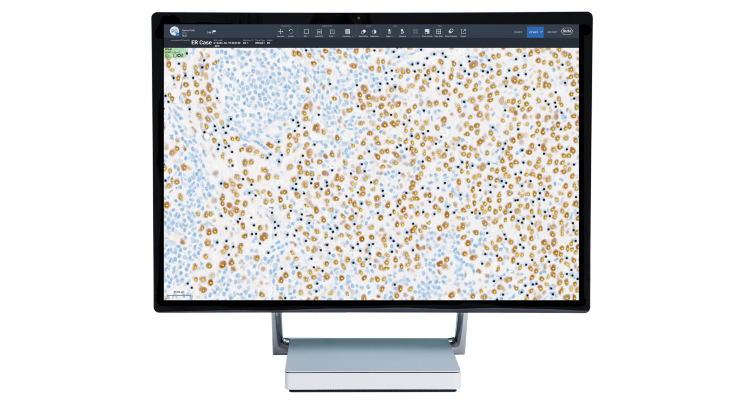
uPath Ki-67 (30-9) image analysis, uPath ER (SP1) image analysis and uPath PR (1E2) image analysis for breast cancer use pathologist-trained deep learning algorithms to enable quick calculation of Ki-67, ER and PR tumor cell nuclei positivity. This includes a whole slide analysis workflow with automated pre-computing of the slide image prior to pathologist assessment, and a clear visual overlay highlighting tumor cells with and without nuclear staining. uPath Ki-67 (30-9) image analysis, uPath ER (SP1) image analysis and uPath PR (1E2) image analysis for breast cancer produce actionable assessments of scanned slide images that are objective and reproducible, aiding pathologists in quantification of these breast cancer markers.
Intended for use with Roche's high medical value assays and slides stained on a BenchMark ULTRA instrument using ultraView DAB detection kit, the uPath Ki-67 (30-9) image analysis, uPath ER (SP1) image analysis and uPath PR (1E2) image analysis algorithms are ready-to-use and integrated within Roche's uPath enterprise software and NAVIFY Digital Pathology, the cloud version of uPath. These algorithms are for Research Use Only. Not for use in diagnostic procedures.
"Roche is committed to the expansion of digital pathology solutions to address unmet medical needs and breast cancer diagnostics is a key opportunity area. Innovations like image analysis algorithms have the potential to impact patient care by increasing the information available to pathologists and enhancing diagnostic confidence," said Jill German, Head of Roche Diagnostics Pathology Customer Area.
In December 2021, Roche will be presenting an abstract (# P1-02-17) on our artificial intelligence, deep learning development of our breast panel RUO algorithms at the San Antonio Breast Cancer Symposium, which features the latest research and development on breast cancer research. To find out more about this symposium, visit https://www.sabcs.org/Symposium-Overview-2021.
References
1 World Health Organization. GLOBOCAN 2020; All cancers fact sheet. [Internet; cited June 2021]. Available from: https://gco.iarc.fr/today/data/factsheets/cancers/39-All-cancers-fact-sheet.pdf
2 Nielsen TO, et al: Assessment of Ki67 in Breast Cancer: Updated Recommendations From the International Ki67 in Breast Cancer Working Group. J Natl Cancer Inst. 2021 Jul 1;113(7):808-819. doi: 10.1093/jnci/djaa201. PMID: 33369635; PMCID: PMC8487652.
