Globe Newswire02.04.19
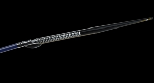
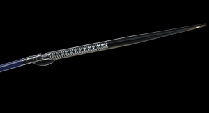
Avinger Inc., a developer of treatments for peripheral artery disease (PAD), has released preliminary analysis of results from its SCAN clinical study, a post-market study comparing optical coherence tomography (OCT) with intravascular ultrasound (IVUS) as a diagnostic imaging tool in the peripheral arteries. Initial analysis of the SCAN data indicated that OCT imaging with Avinger’s Pantheris System showed statistical superiority or equivalence to IVUS on all parameters evaluated.
Avinger had previously announced completion of enrollment in the SCAN study in August 2018. During the study, 120 OCT and IVUS matched images (240 images in total) were captured at identical positions within vessel segments from 12 subjects at two clinical centers. Three independent radiologists then reviewed the matched images and ranked them in terms of image quality and ability to display vessel morphology and disease. Vessel measurement capabilities were also compared between the two imaging modalities.
The preliminary data analysis from the reviewer rankings of the 240 images indicated that OCT imaging with Pantheris was significantly better than IVUS in visualizing plaque, calcification and stent struts. OCT imaging was ranked to be statistically equivalent to IVUS in visualizing the layered structures of the vessel wall and visualizing troughs following atherectomy. Vessel measurement and interference of artifacts with imaging quality were also seen to be statistically equivalent between the two imaging modalities. It is anticipated that final SCAN study results will be submitted for publication by its physician investigators, Dr. Edward Pavillard, a vascular surgeon at PA Vascular Institute in King of Prussia, Pa., and Dr. Luke Sewall, an interventional radiologist and president of Vascular and Interventional Professionals in Hinsdale, Ill.
Dr. Pavillard commented, “The SCAN study does an excellent job demonstrating the clinical value of intravascular imaging with OCT and IVUS for the diagnosis of peripheral arterial disease. While OCT performed better on certain parameters, what is paramount to providing excellent clinical results is the capability to see what is present in the vessel so that diseased tissue can be removed without causing injury to healthy tissue. To our knowledge, SCAN is only the second study in the world and the first U.S.-based study comparing OCT with IVUS intravascular imaging in the peripheral vessels. We look forward to sharing more detailed findings of the study as they become available.”
Avinger’s proprietary Lumivascular technology utilizes OCT to provide real-time vascular imaging for the diagnosis and treatment of peripheral artery disease. OCT is a light-based, high-definition imaging modality, which generates no X-Ray radiation, and has a resolution approximately ten times greater than IVUS. The U.S. Food and Drug Administration (FDA) has cleared both diagnostic and therapeutic treatment indications for use for Avinger’s Pantheris (atherectomy) and Ocelot (CTO-crossing) image-guided catheters. While reimbursement codes applicable to therapeutic peripheral interventions with the Company’s image-guided catheters already exist, the results of the SCAN study are intended to support an initiative to gain incremental reimbursement for OCT diagnostic imaging in the peripheral arteries similar to the reimbursement currently provided for the use of IVUS as a diagnostic tool in this setting.
Avinger is a commercial-stage medical device company that designs and develops the first-ever image-guided, catheter-based system that diagnoses and treats patients with peripheral artery disease (PAD). PAD is estimated to affect over 12 million people in the United States and over 200 million worldwide. Avinger is dedicated to radically changing the way vascular disease is treated through its Lumivascular platform, which currently consists of the Lightbox imaging console, the Ocelot family of chronic total occlusion (CTO) catheters, and the Pantheris family of atherectomy devices. Avinger is based in Redwood City, Calif.