University of Glasgow12.22.15
Researchers from the University of Glasgow have found a way to make swallowable cameras more effective at detecting cancers of the throat and gut.
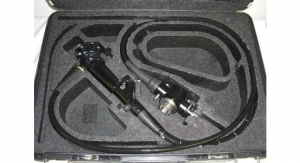
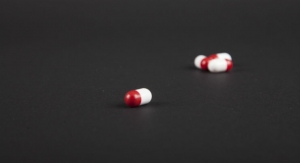
In recent years, tiny sensing systems small enough for patients to swallow have proven to be a valuable clinical alternative to more intrusive imaging methods such as endoscopes.
Until now, the systems, often known as video-pill, have relied on illuminating patients’ innards using a small light source, restricting clinicians to conclusions based on what they can see in the spectrum of visible light.
In a new paper published today (Friday, December 18) in the journal Scientific Reports, researchers from the University’s School of Engineering describe how they have used fluorescent light for the first time to expand the diagnostic capabilities of the video-pill.
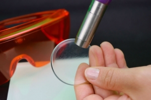
Fluorescence imaging is already a powerful diagnostic tool in medicine, capable of clearly identifying in patients the rich blood supplies which support cancers and help them to grow, but which can be missed by examination under visible light. However, past fluorescence imaging technologies have been expensive, bulky and consume substantial power, confining the technique to laboratories and hospital examination rooms.
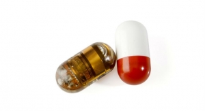
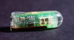
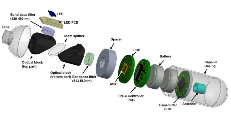
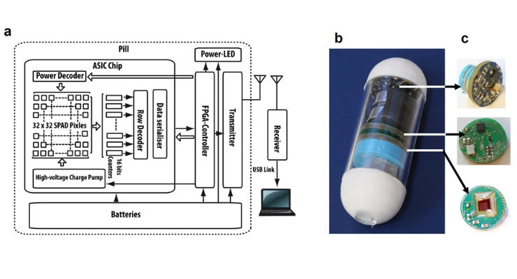
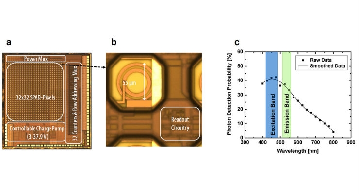
Using an advanced semiconductor single-pixel imaging technique, the researchers have managed to create flurorescence imaging in a small pill form for the first time.
David Cumming, a professor in Electronic and Nanoscale Engineering at the University of Glasgow, led the project.
“The system we’ve developed is small enough and power efficient enough to image the entire human gastrointestinal tract for up to 14 hours,” said research associate Dr. Mohammed Al-Rawhani. “We’ve confirmed in the lab the ability of the system to image fluorescence ‘phantoms’—mixtures of flavin and hemoglobin, which mimic closely how cancers are affected by fluorescence in parts of the body like the intestines, the bowel, and the esophagus.
“The system could also be used to help track antibodies used to label cancer in the human body, creating a new way to detect of cancer,” added Dr. Al-Rawhani. “It’s a valuable new technique, which could help clinicians make fewer false positives and negatives in cancer diagnosis, which could lead to more effective treatment in the future.”
“We’ve played an important role in developing the technology behind video-pill systems, and this is an exciting new development, which offers a valuable new resource for gastrointestinal imaging,” said Cumming. “There’s still some way to go before it will be ready for commercial production and clinical use, but we’re in early talks with industry to bring a product to market. We’re also interested in expanding the imaging capabilities of video-pill systems to new areas such as ultrasound in the near future.”
The paper, titled ‘Wireless fluorescence capsule for endoscopy using single photon-based detection’ is published in Scientific Reports.
In recent years, tiny sensing systems small enough for patients to swallow have proven to be a valuable clinical alternative to more intrusive imaging methods such as endoscopes.
Until now, the systems, often known as video-pill, have relied on illuminating patients’ innards using a small light source, restricting clinicians to conclusions based on what they can see in the spectrum of visible light.
In a new paper published today (Friday, December 18) in the journal Scientific Reports, researchers from the University’s School of Engineering describe how they have used fluorescent light for the first time to expand the diagnostic capabilities of the video-pill.
Fluorescence imaging is already a powerful diagnostic tool in medicine, capable of clearly identifying in patients the rich blood supplies which support cancers and help them to grow, but which can be missed by examination under visible light. However, past fluorescence imaging technologies have been expensive, bulky and consume substantial power, confining the technique to laboratories and hospital examination rooms.
Using an advanced semiconductor single-pixel imaging technique, the researchers have managed to create flurorescence imaging in a small pill form for the first time.
David Cumming, a professor in Electronic and Nanoscale Engineering at the University of Glasgow, led the project.
“The system we’ve developed is small enough and power efficient enough to image the entire human gastrointestinal tract for up to 14 hours,” said research associate Dr. Mohammed Al-Rawhani. “We’ve confirmed in the lab the ability of the system to image fluorescence ‘phantoms’—mixtures of flavin and hemoglobin, which mimic closely how cancers are affected by fluorescence in parts of the body like the intestines, the bowel, and the esophagus.
“The system could also be used to help track antibodies used to label cancer in the human body, creating a new way to detect of cancer,” added Dr. Al-Rawhani. “It’s a valuable new technique, which could help clinicians make fewer false positives and negatives in cancer diagnosis, which could lead to more effective treatment in the future.”
“We’ve played an important role in developing the technology behind video-pill systems, and this is an exciting new development, which offers a valuable new resource for gastrointestinal imaging,” said Cumming. “There’s still some way to go before it will be ready for commercial production and clinical use, but we’re in early talks with industry to bring a product to market. We’re also interested in expanding the imaging capabilities of video-pill systems to new areas such as ultrasound in the near future.”
The paper, titled ‘Wireless fluorescence capsule for endoscopy using single photon-based detection’ is published in Scientific Reports.