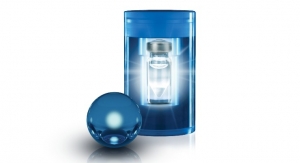
The probe is approximately one-hundredth the width of a human hair and is composed of two magnetic disks attached by a hydrogel spacer that is designed to expand or contract in response to various conditions within the body. As the hydrogel changes shape, the magnetic fields surrounding the disks become weaker or stronger, changing the frequency with which the water molecule protons in the gel resonate in response to applied RF radiation.
Between the disks is a spacer layer of hydrogel, a polymer network that can absorb water and expand significantly; the amount of expansion depends on the chemical properties of the gel and the environment around it. Conversely, it also can shrink in response to changing local conditions. Swelling or shrinking of the gel changes the distance (and hence, the magnetic field strength) between the two disks, and that, in turn, changes the frequency at which the protons in water molecules around and inside the gel resonate in response to radio-frequency radiation. Scanning the sample with a range of frequencies quickly identifies the current shape of the nanoprobes, effectively measuring the remote conditions through the changes in resonance frequencies caused by the shape-changing agents.
In the experiments reported in the journal Nature, the scientists tested the sensors in solutions of varying pH, in solutions with ion concentration gradients, and in a liquid growth medium containing living canine kidney cells as their metabolism went from normal to nonfunctional in the absence of oxygen. That phenomenon caused the growth medium to acidify, and the change over time was sensed by the GEMs and recorded through real-time shifting in resonant frequencies. Even for the un-optimized, first-generation probes used, the frequency shifts resulting from changes in pH were easily resolvable and orders of magnitude larger than any equivalent frequency shifting observed through traditional magnetic resonance spectroscopy approaches.
Tracking highly localized pH values in living organisms can be difficult. (A blood test cannot necessarily do it because the sample mixes blood from numerous locations.) Yet local pH changes can provide invaluable early signals of many pathologies. For example, the pH around a cancer cell is slightly lower than normal, and internal inflammation generally leads to local change in pH level. Detecting such changes might reveal, for example, the presence of an unseen tumor or show whether an infection has developed around a surgical implant.
"Of course, that sort of potential use in living organisms is still a long way off," said Gary Zabow, a NIST scientist and team leader. "Our data were taken in-vitro. And some potential applications of the sensors may not be biological at all. But a long-term goal is to improve our techniques to the point at which GEMs can be employed for biomedical uses."
That would require, among other things, further miniaturization. The 0.5 to 2 µm diameter GEMs in the experiments are already small enough for many in-vitro and other possible non-biological applications, as well as possibly for some in-vivo cellular related applications. But preliminary estimates by the experimenters indicate that the sensors can be reduced substantially from their current size, and might conceivably be made smaller than 100 nanometers in diameter. That would open up many additional biomedical applications.
One of the most significant features of GEMs is that they can be "tuned" in fabrication to respond to different biochemical states and to resonate in different parts of the RF spectrum by altering the gel composition and the magnet shapes and materials, respectively. So placing two different populations of GEMs at the same site makes it possible to track changes in two different variables at the same time—a capability the researchers demonstrated by placing GEMs with two different dimensions in the same location and detecting the signals from both simultaneously.
"The idea is that you could design different sensors to measure different things, effectively measuring a panel of potential biomarkers simultaneously, rather than just one, to better differentiate between different pathologies," Zabow said. "We think that these sensors can potentially be adapted to measure a variety of different biomarkers, possibly including things such as glucose, local temperatures, various ion concentrations, possibly the presence or absence of various enzymes and so forth."
Ron Goldfarb, leader of NIST's Magnetics Group, noted that, "the work on geometrically encoded magnetic sensors by Gary Zabow and colleagues is a natural extension of research published by the team, along with NIST's John Moreland, in 2008. That work showed how micromagnets can act as 'smart tags' to potentially identify particular cells, tissues or physiological conditions. Functionally, the GEMS in the current effort are more advanced in that they change their shape in response to stimuli; thus, they act as measurement devices. The next challenge will be design optimization and the development of dimensionally controlled, large-scale fabrication processes in order to make these sensors widely available to researchers."