Rachel Klemovitch, Assistant Editor04.29.24
Through a new study at the Tokyo University of Science, researchers have developed the world’s first rigid endoscope for visible-to-OTN HSI, utilizing supercontinuum lighting and an acoustic-opto tunable filter. This system has the potential to lead the way for advanced medical imaging, helping both physicians and patients.
Hyperspectral imaging (HSI) is a state-of-the-art technique that captures and processes information across a given electromagnetic spectrum and is useful for determining the composition of a variety of objects. Unlike traditional imaging techniques that capture light intensity at specific wavelengths, HSI collects a full spectrum at each pixel in an image. This distinguishes different materials and substances based on their unique spectral signatures.
Near-infrared hyperspectral imaging (NIR-HSI) has over-thousand-nanometer (OTN) spectroscopy, which can be used for the identification of organic substances, their concentration estimation, and 2D map creation. NIR-HSI can also be used to gain information deep into the body, making it useful for the visualization of lesions hidden in normal tissues.
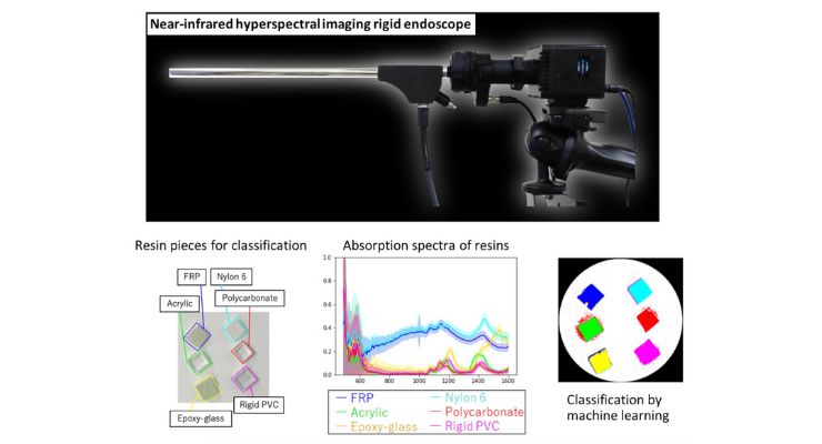
The new study led by Professor Hiroshi Takemura from Tokyo University of Science (TUS) and Toshihiro Takamatsu, Ryodai Fukushima, Kounosuke Sato, Masakazu Umezawa, and Kohei Soga, all from TUS; Hideo Yokota from RIKEN, and Abian Hernandez Guedes and Gustavo M. Calico, from the University of Las Palmas de Gran Canaria, has recently developed the world’s first rigid endoscope system capable of HSI from visible to OTN wavelengths.
Researchers used a supercontinuum (SC) light source and an acoustic-opto tunable filter (AOTF) that can emit specific wavelengths. The optical performance and classification were verified by the team, demonstrating its capability to perform HSI in the range of 490–1600 nm, enabling visible as well as NIR-HSI.
Results also showed the system’s advantages such as enabling non-destructive imaging, the low light power of extracted wavelengths, and downsizing capabilities. a more continuous NIR spectrum can be obtained compared to that of conventional rigid-scope-type devices.
Prof. Takemura explained, “An SC light source can output intense coherent white light, whereas an AOTF can extract light containing a specific wavelength. This combination offers easy light transmission to the light guide and the ability to electrically switch between a broad range of wavelengths within a millisecond.”
Researchers used the system to acquire the spectra of six types of resins and employed a neural network to classify the spectra pixel-by-pixel in multiple wavelengths. Results showed that when the OTN wavelength range was extracted from the HSI data for training, the neural network could classify seven different targets, including the six resins and a white reference, with an accuracy of 99.6%, reproducibility of 93.7%, and specificity of 99.1%. This means that the system can successfully extract molecular vibration information of each resin at each pixel.
Results were published in Optics Express on April 17, 2024.
Prof. Takemura added: “This breakthrough, which combines expertise from different fields through a collaborative, cross-disciplinary approach, enables the identification of invaded cancer areas and the visualization of deep tissues such as blood vessels, nerves, and ureters during medical procedures, leading to improved surgical navigation. Additionally, it enables measurement using light previously unseen in industrial applications, potentially creating new areas of non-use and non-destructive testing. By visualizing the invisible, we aim to accelerate the development of medicine and improve the quality of life of physicians as well as patients.”
Hyperspectral imaging (HSI) is a state-of-the-art technique that captures and processes information across a given electromagnetic spectrum and is useful for determining the composition of a variety of objects. Unlike traditional imaging techniques that capture light intensity at specific wavelengths, HSI collects a full spectrum at each pixel in an image. This distinguishes different materials and substances based on their unique spectral signatures.
Near-infrared hyperspectral imaging (NIR-HSI) has over-thousand-nanometer (OTN) spectroscopy, which can be used for the identification of organic substances, their concentration estimation, and 2D map creation. NIR-HSI can also be used to gain information deep into the body, making it useful for the visualization of lesions hidden in normal tissues.
The new study led by Professor Hiroshi Takemura from Tokyo University of Science (TUS) and Toshihiro Takamatsu, Ryodai Fukushima, Kounosuke Sato, Masakazu Umezawa, and Kohei Soga, all from TUS; Hideo Yokota from RIKEN, and Abian Hernandez Guedes and Gustavo M. Calico, from the University of Las Palmas de Gran Canaria, has recently developed the world’s first rigid endoscope system capable of HSI from visible to OTN wavelengths.
Researchers used a supercontinuum (SC) light source and an acoustic-opto tunable filter (AOTF) that can emit specific wavelengths. The optical performance and classification were verified by the team, demonstrating its capability to perform HSI in the range of 490–1600 nm, enabling visible as well as NIR-HSI.
Results also showed the system’s advantages such as enabling non-destructive imaging, the low light power of extracted wavelengths, and downsizing capabilities. a more continuous NIR spectrum can be obtained compared to that of conventional rigid-scope-type devices.
Prof. Takemura explained, “An SC light source can output intense coherent white light, whereas an AOTF can extract light containing a specific wavelength. This combination offers easy light transmission to the light guide and the ability to electrically switch between a broad range of wavelengths within a millisecond.”
Researchers used the system to acquire the spectra of six types of resins and employed a neural network to classify the spectra pixel-by-pixel in multiple wavelengths. Results showed that when the OTN wavelength range was extracted from the HSI data for training, the neural network could classify seven different targets, including the six resins and a white reference, with an accuracy of 99.6%, reproducibility of 93.7%, and specificity of 99.1%. This means that the system can successfully extract molecular vibration information of each resin at each pixel.
Results were published in Optics Express on April 17, 2024.
Prof. Takemura added: “This breakthrough, which combines expertise from different fields through a collaborative, cross-disciplinary approach, enables the identification of invaded cancer areas and the visualization of deep tissues such as blood vessels, nerves, and ureters during medical procedures, leading to improved surgical navigation. Additionally, it enables measurement using light previously unseen in industrial applications, potentially creating new areas of non-use and non-destructive testing. By visualizing the invisible, we aim to accelerate the development of medicine and improve the quality of life of physicians as well as patients.”
