09.02.15
Modification of atherosclerotic plaque with the Diamondback 360 orbital atherectomy device (Cardiovascular Systems Inc.) improved drug uptake in calcified peripheral lesions from human cadavers, according to recent study results.
“We know from clinical studies that anti-restenotic drug effects are diminished in severely calcified arteries,” said Gunnar Tepe, M.D., with RoMed Hospital in Rosenheim, Germany.
“The data are exciting in that they suggest that subtle modification to the plaque surface can have profound effects on drug penetration. Massive debulking may be a relic of the past, and a more muted approach may extend endovascular intervention for PAD treatment into vessels even below the knee,” said Elazer Edelman, M.D., Ph.D., chairman and co-founder of CBSET, and a co-author of the study.
In Europe and North America, an estimated 27 million individuals are affected with peripheral artery disease (PAD), which is caused by the accumulation of plaque in peripheral arteries (commonly the pelvis or leg) reducing blood flow. Left untreated, PAD can lead to severe pain, immobility, non-healing wounds and eventually limb amputation. With risk factors such as diabetes and obesity on the rise, the prevalence of PAD is growing at double-digit rates.
“A more sophisticated understanding of drug transfer after plaque modification is critically important to optimizing PAD therapies for improved clinical outcomes,” said Peter Markham, president/CEO and a co-founder of CBSET.
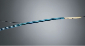
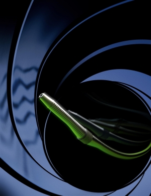
Millions of patients with PAD may benefit from treatment with orbital atherectomy utilizing Cardiovascular System Inc.’s (CSI) peripheral orbital atherectomy systems, minimally invasive catheter systems developed and manufactured by CSI. These systems use a diamond-coated crown, attached to an orbiting shaft, which targets atherosclerotic plaque while preserving healthy vessel tissue — a critical factor in preventing reoccurrences. Balloon angioplasty and stents have significant shortcomings in treating hard, calcified lesions. Stents are prone to fractures and high recurrence rates, and treatment of hard, calcified lesions often leads to vessel damage and suboptimal results.
“This study is part of a larger effort by CBSET, MIT and collaborating companies to leverage benchtop, animal, and computational studies in order to define the barriers to drug distribution in healthy and diseased arteries,” explained Rami Tzafriri, Ph.D., principal scientist at CBSET. “While these initial findings illustrate, for the first time, the barrier effects of calcified plaque on arterial drug diffusion, they provide a path for improved therapy, as even relatively benign modification of this plaque barrier can profoundly improve drug delivery into diseased peripheral arteries.”
Based in Lexington, Mass., CBSET is a non-profit preclinical research institute dedicated to biomedical research, education, and advancement of medical technologies. CBSET offers the latest equipment for fluoroscopy, echocardiography (TEE/TTE), electrophysiology, IVUS, optical coherence tomography (OCT), endoscopy/laparoscopy, surgical video recording, histology, microradiography, and SEM (scanning electron microscopy).
“We know from clinical studies that anti-restenotic drug effects are diminished in severely calcified arteries,” said Gunnar Tepe, M.D., with RoMed Hospital in Rosenheim, Germany.
“The data are exciting in that they suggest that subtle modification to the plaque surface can have profound effects on drug penetration. Massive debulking may be a relic of the past, and a more muted approach may extend endovascular intervention for PAD treatment into vessels even below the knee,” said Elazer Edelman, M.D., Ph.D., chairman and co-founder of CBSET, and a co-author of the study.
In Europe and North America, an estimated 27 million individuals are affected with peripheral artery disease (PAD), which is caused by the accumulation of plaque in peripheral arteries (commonly the pelvis or leg) reducing blood flow. Left untreated, PAD can lead to severe pain, immobility, non-healing wounds and eventually limb amputation. With risk factors such as diabetes and obesity on the rise, the prevalence of PAD is growing at double-digit rates.
“A more sophisticated understanding of drug transfer after plaque modification is critically important to optimizing PAD therapies for improved clinical outcomes,” said Peter Markham, president/CEO and a co-founder of CBSET.
Millions of patients with PAD may benefit from treatment with orbital atherectomy utilizing Cardiovascular System Inc.’s (CSI) peripheral orbital atherectomy systems, minimally invasive catheter systems developed and manufactured by CSI. These systems use a diamond-coated crown, attached to an orbiting shaft, which targets atherosclerotic plaque while preserving healthy vessel tissue — a critical factor in preventing reoccurrences. Balloon angioplasty and stents have significant shortcomings in treating hard, calcified lesions. Stents are prone to fractures and high recurrence rates, and treatment of hard, calcified lesions often leads to vessel damage and suboptimal results.
“This study is part of a larger effort by CBSET, MIT and collaborating companies to leverage benchtop, animal, and computational studies in order to define the barriers to drug distribution in healthy and diseased arteries,” explained Rami Tzafriri, Ph.D., principal scientist at CBSET. “While these initial findings illustrate, for the first time, the barrier effects of calcified plaque on arterial drug diffusion, they provide a path for improved therapy, as even relatively benign modification of this plaque barrier can profoundly improve drug delivery into diseased peripheral arteries.”
Based in Lexington, Mass., CBSET is a non-profit preclinical research institute dedicated to biomedical research, education, and advancement of medical technologies. CBSET offers the latest equipment for fluoroscopy, echocardiography (TEE/TTE), electrophysiology, IVUS, optical coherence tomography (OCT), endoscopy/laparoscopy, surgical video recording, histology, microradiography, and SEM (scanning electron microscopy).