Previously, doctors were able to study patient anatomy only by mentally putting together information from multiple screens, said EchoPixel’s CEO Ron Schilly. The use of 2-D digital scans revolutionized medicine, because they allowed doctors to see an individual patient's anatomy without cutting into the body. "But when a doctor evaluates them, they are looking at a series of 2-D slices and trying to create that 3-D anatomy in their mind,” Sergio Aguirre, founder and CTO of EchoPixel, told Smithsonian.com. “Doctors are focusing energy on solving this 3-D problem instead of the clinical problem at hand, and we think this software will help them get a clearer view of the problem more quickly.”
Other systems, like GE's Vivid E9 with XDclear, already compile such images to produce 3-D visuals that appear very realistic, and they even have 3-D properties that allow them to be rotated or taken apart. But they remain limited to display on a flat screen. EchoPixel appears to take 3-D imaging a step further by generating interactive holograms.
Experts have become very good at reading 2-D images and manipulating 3-D representations on a flat screen, so holograms may not add a huge advantage in some applications, Sandy Napel, co-director of Stanford University's Radiology 3-D and Quantitative Imaging Laboratory, noted to Smithsonian.com. But there are specific procedures that EchoPixel may be primed to improve. For instance, the technology is already being tested at the University of California, San Francisco, for virtual colonoscopies—an alternative to the unpopular procedure in which a colonoscope is inserted and manipulated within the human body.
True 3-D medical imaging also may benefit doctors who need to visualize abnormal or complex 3-D structures, like the mess of broken and displaced bones that can result from trauma due to a motor vehicle accident. “A surgeon that's going to plan to remove fragments and fix that kind of injury might benefit by viewing a true 3-D representation of what they will actually see when they have the patient in the operating room,” Napel said. “I think having, say, a 3-D pelvis floating above a desktop, where you could see all the actual fractures and displacements, could have great potential for surgical planning.”Heart conditions in very young patients are another area where medical holograms could shine. “The heart is a complicated structure, but every medical student can draw a picture of a normal heart,” Napel said. “However, when you have narrowings, aneurisms, congenital abnormalities—being able to visualize these in 3-D could be really helpful. Think about kids born with genetic defects that cause the heart to develop abnormally. A surgeon is going to go in there and operate on a very young human being and hopefully make a correction. They'll get a careful report from a radiologist, saying that some blood vessels are connected here and they should be there, and the surgeons can see it also on CT scans, but not the same way they are going to see it in the operating room." Having a 3D preview of what they'll see when they start surgery could help the doctor understand the situation much more quickly.
Early clinical trials have shown True3D technology to impact procedures in surgery and radiology, a news release said. EchoPixel uses proprietary protocols to formulate and distribute expert-derived methodologies in step-by-step 3-D format, according to the release.EchoPixel is a privately held, venture backed company located at the Fogarty Institute for Innovation in Mountain View, Calif.
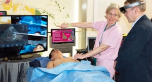
True3D compiles data from MRI, CT, and all DICOM imaging data to create an interactive holograph of patient-specific body parts, instead of flat 2D images, EchoPixel said in a release. The rendering allows physicians and students to interact with the anatomical structures. True3D’s technology is an effort to dramatically improve speed and efficacy across a broad range of medical operations, the release said.
“Since CT scans were invented in the 1970s, doctors have learned about patient anatomy by mentally piecing together multiple images on flat screens,” Ron Schilling, CEO of EchoPixel, said in the release. “That’s not what the inside of a patient looks like. When working with doctors, we found they were wasting energy trying to solve imaging problems instead of clinical ones. Using virtual reality, we can provide an interactive, three-dimensional view of patient data that is far clearer and more realistic.”
Early clinical trials have shown True3D technology to impact procedures in surgery and radiology, the release said. EchoPixel uses proprietary protocols to formulate and distribute expert-derived methodologies in step-by-step 3D format, according to the release.
- See more at: http://www.diagnosticimaging.com/ct/echopixel-launches-3d-virtual-imaging-fda-nod-0#sthash.8f8giqbr.dpuf